. 2024 Jan 16;15(1):e0292423.
doi: 10.1128/mbio.02924-23. Epub 2023 Dec 7.

Bacteriophage infection and killing of intracellular Mycobacterium abscessus

Affiliations

Bacteriophage infection and killing of intracellular Mycobacterium abscessus

Alan A Schmalstig et al. mBio. .

Abstract

As we rapidly approach a post-antibiotic era, bacteriophage (phage) therapy may offer a solution for treating drug-resistant bacteria. Mycobacterium abscessus is an emerging, multidrug-resistant pathogen that causes disease in people with cystic fibrosis, chronic obstructive pulmonary disease, and other underlying lung diseases. M. abscessus can survive inside host cells, a niche that can limit access to antibiotics. As current treatment options for M. abscessus infections often fail, there is an urgent need for alternative therapies. Phage therapy is being used to treat M. abscessus infections as an option of last resort. However, little is known about the ability of phages to kill bacteria in the host environment and specifically in an intracellular environment. Here, we demonstrate the ability of phages to enter mammalian cells and to infect and kill intracellular M. abscessus. These findings support the use of phages to treat intracellular bacterial pathogens.

Keywords: Mycobacterium abscessus; bacteriophage therapy; intracellular bacteria; macrophages; nontuberculous mycobacteria.


Conflict of interest statement

The authors declare no conflict of interest.

Figures

Fig 1
Fig 1
Phage sensitivity of specific mycobacterial strains. Serially diluted phages were spotted on lawns of M. smegmatis mc2155, M. abscessus GD82, or M. abscessus GD20. Cleared spots in the lawn indicate phage-mediated killing.
Fig 2
Fig 2
Clinical M. abscessus strains grow in mammalian cells. Mammalian cells were infected with M. abscessus strains at an MOI of 10. (A) THP-1 cells infected with M. abscessus GD82. (B) THP-1 cells infected with M. abscessus GD20 (blue) and M. abscessus ATCC 19977 Rough (black). (C) A549 cells infected with GD82. (D) Murine BMDM infected with GD82. Triplicate wells of infected mammalian cells were lysed for each time point between 0 and 5 days post infection and plated for CFU enumeration. Data shown are representative of two independent experiments. **P < 0.01, ***P < 0.001, ****P < 0.0001 by one-way ANOVA with Tukey’s post hoc test when compared to day 0.
Fig 3
Fig 3
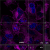
SYBR Gold phage are internalized by A549 cells. A 3D deconvoluted Z-series was used to generate an orthogonal view of BPsΔ (MOI 105) inside A549 cells. Phage were stained with SYBR Gold (green). A549 cells were stained with CellMask plasma membrane (magenta) and DAPI (blue). Side views represent the Z dimension. Scale bar is 10 µm.
Fig 4
Fig 4
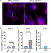
Mammalian cells internalize phage. Phages were stained with SYBR Gold and incubated with mammalian cells for 24 h. (A) Representative images of intracellular phage (green) at an MOI of 103. Mammalian cells were stained with CellMask plasma membrane (magenta) and DAPI (blue). White arrows indicate mammalian cells with intracellular phage puncta. Scale bar is 10 µm. Percentage of THP-1 cells (B), BMDMs (C), and A549 cells (D) with intracellular phage. For B and C, phage were added to mammalian cells at an MOI of 103. For D, phage were added at an MOI of 103 or 105. Error bars represent standard deviation from two independent experiments, each with a minimum of 14 fields of view counted. A minimum of 300 mammalian cells were counted per experiment. ns, not significant; *P < 0.05, ***P < 0.001, ****P < 0.0001 by Kruskal–Wallis one-way ANOVA with Dunn’s post hoc test.
Fig 5
Fig 5
mCherry reporter phages infect intracellular mycobacteria. (A) Representative images of mCherry reporter phage infection of intracellular GD82 in A549 cells. A549 cells were infected with GFP-expressing GD82 at an MOI of 10, washed to remove extracellular bacteria, infected with BPs∆::mCherry or ZoeJΔ::mCherry phage at an MOI of 104, and imaged after 24 h. Mammalian cells were stained with CellMask plasma membrane (magenta) and DAPI (blue). Scale bar is 10 µm. Red bacilli indicate phage-infected M. abscessus. Percent reporter phage infection of intracellular M. abscessus GD82 in THP-1 cells (B), GD82 in A549 cells (C), and GD20 in THP-1 cells (D). Error bars represent standard deviation from two independent experiments each with a minimum of 12 fields of view counted per experiment. A minimum of 350 M. abscessus cells (GFP) were counted per experiment. ns, not significant; ****P < 0.0001 by Mann–Whitney test.
Fig 6
Fig 6
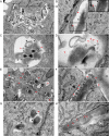
Transmission electron microscopy of phage infection of intracellular M. abscessus. GD20-infected THP-1 cells were incubated with BPsΔ for 24 h (A–D). THP-1 phagosome with BPsΔ phage adsorbed to intracellular GD20 (A and B). BPsΔ phage tails adsorb to intracellular GD20 (C). BPsΔ phage, with empty and intact capsids, adsorbed to intracellular GD20 (D). GD82-infected THP-1 cells were incubated with BPsΔ for 24 h. Colocalized phage and bacteria are observed in multiple phagosomes in the same cell (E). GD82-infected THP-1 cells incubated with BPsΔ for 48 h. Multiple phages adsorbed to the bacterial pole are observed (F). GD82-infected A549 cells incubated with BPsΔ for 24 h and a phage tail adsorbed to intracellular bacteria (G). GD82-infected A549 cells were incubated with BPsΔ for 48 h, and intracellular phage progeny was observed (H). Black arrowheads indicate intracellular M. abscessus, red arrowheads indicate adsorbed phage, black arrows indicate phage tails, red outlined arrowheads indicate empty phage capsid, and red arrows indicate phage progeny. Only a subset of phage particles and bacteria are indicated. Black boxes indicate areas of higher magnification (A and B). For additional TEM images, see Fig. S5.
Fig 7
Fig 7
Phages kill intracellular M. abscessus GD82 in mammalian cells. (A) THP-1 cells, (B) BMDM, or (C) A549 cells were infected with M. abscessus GD82 at an MOI of 10, washed to remove extracellular GD82, and then treated with BPsΔ, ZoeJΔ, or Muddy. After 48 h, mammalian cells were lysed in the presence of PIB and plated for bacterial CFU enumeration. Error bars represent standard deviation from a minimum of two independent experiments determined by one-way ANOVA with Tukey’s post hoc test; ns, not significant; *P < 0.05, **P < 0.01, ***P < 0.001, ****P < 0.0001.

References

    1. Projan SJ. 2003. Why is big Pharma getting out of antibacterial drug discovery? Curr Opin Microbiol 6:427–430. doi:10.1016/j.mib.2003.08.003 - DOI - PubMed
    1. Chokshi A, Sifri Z, Cennimo D, Horng H. 2019. Global contributors to antibiotic resistance. J Glob Infect Dis 11:36–42. doi:10.4103/jgid.jgid_110_18 - DOI - PMC - PubMed
    1. Magiorakos A-P, Srinivasan A, Carey RB, Carmeli Y, Falagas ME, Giske CG, Harbarth S, Hindler JF, Kahlmeter G, Olsson-Liljequist B, Paterson DL, Rice LB, Stelling J, Struelens MJ, Vatopoulos A, Weber JT, Monnet DL. 2012. Multidrug-resistant, extensively drug-resistant and pandrug-resistant bacteria: an international expert proposal for interim standard definitions for acquired resistance. Clin Microbiol Infect 18:268–281. doi:10.1111/j.1469-0691.2011.03570.x - DOI - PubMed
    1. Beyer P, Paulin S. 2020. Priority pathogens and the antibiotic pipeline: an update. Bull World Health Organ 98:151. doi:10.2471/BLT.20.251751 - DOI - PMC - PubMed
    1. Luepke KH, Suda KJ, Boucher H, Russo RL, Bonney MW, Hunt TD, Mohr III JF. 2017. Past, present, and future of antibacterial economics: increasing bacterial resistance, limited antibiotic pipeline, and societal implications. Pharmacotherapy 37:71–84. doi:10.1002/phar.1868 - DOI - PubMed

Substances

LinkOut - more resources