. 2024 Jul 1;16(1):mjad079.
doi: 10.1093/jmcb/mjad079.

Pathologically relevant aldoses and environmental aldehydes cause cilium disassembly via formyl group-mediated mechanisms

Affiliations

Pathologically relevant aldoses and environmental aldehydes cause cilium disassembly via formyl group-mediated mechanisms

Te Li et al. J Mol Cell Biol. .

Abstract

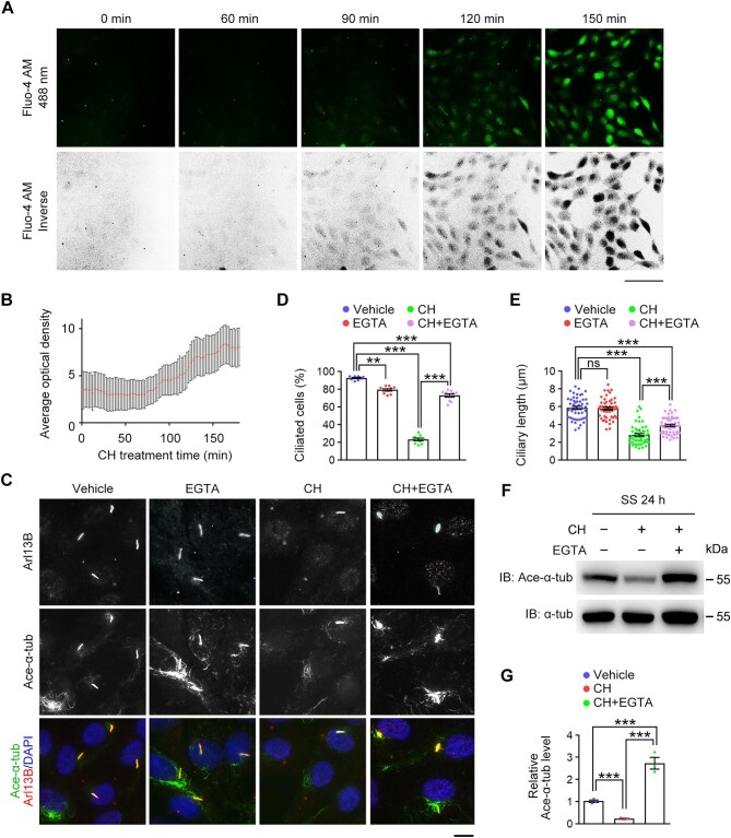
Carbohydrate metabolism disorders (CMDs), such as diabetes, galactosemia, and mannosidosis, cause ciliopathy-like multiorgan defects. However, the mechanistic link of cilia to CMD complications is still poorly understood. Herein, we describe significant cilium disassembly upon treatment of cells with pathologically relevant aldoses rather than the corresponding sugar alcohols. Moreover, environmental aldehydes are able to trigger cilium disassembly by the steric hindrance effect of their formyl groups. Mechanistic studies reveal that aldehydes stimulate extracellular calcium influx across the plasma membrane, which subsequently activates the calmodulin-Aurora A-histone deacetylase 6 pathway to deacetylate axonemal microtubules and triggers cilium disassembly. In vivo experiments further show that Hdac6 knockout mice are resistant to aldehyde-induced disassembly of tracheal cilia and sperm flagella. These findings reveal a previously unrecognized role for formyl group-mediated cilium disassembly in the complications of CMDs.

Keywords: HDAC6; aldehyde; aldose; calcium influx; carbohydrate metabolism disorder; cilium disassembly; formyl group.


Figures

Figure 1
Figure 1
Aldoses trigger cilium disassembly in a formyl group-dependent manner. (A) Strategy used to study the effects of aldoses on cilia. Ciliated RPE-1 cells were incubated with aldoses for 12 h, followed by quantitative analysis for the percentage of ciliated cells and ciliary length. (BK) RPE-1 cells were incubated with vehicle (PBS) or the indicated compounds (25 mM) in serum-free medium. (B, D, F, and J) Immunofluorescence images of ciliated cells stained for Arl13B, acetylated α-tubulin, and 4′,6-diamidino-2-phenylindole(DAPI). Scale bar, 10 μm. (C, E, G, and K) The percentage of ciliated cells (n = 10 fields from three independent experiments) and quantification of ciliary length (n = 50 cilia from three independent experiments). (H and I) Immunoblotting for O-GlcNAc, O-GlcNAc transferase (OGT), and αtubulin. Data are presented as mean ± SEM. An unpaired two-tailed t-test was performed. ***P < 0.001; ns, not significant.
Figure 2
Figure 2
The cilium disassembly activity of aldehydes is regulated by steric hindrance of the formyl groups. (A) Chemical structures of the aldehydes used in this study. FA, formaldehyde; AH, acetaldehyde; PPA, propionaldehyde; BA, butyraldehyde; VA, valeraldehyde; BZA, benzaldehyde. (B and C) RPE-1 cells were incubated with vehicle (DMSO) or the indicated aldehydes in serum-free medium. (B) Immunofluorescence images of ciliated cells stained for γ-tubulin, acetylated α-tubulin, and DAPI. Scale bar, 10 μm. (C) Quantification of the percentage of ciliated cells (n = 10 fields from 3 independent experiments). (D) Chemical structures of trichloroacetaldehyde (TCAA) and its analogues trifluoroacetaldehyde (TFAA) and monochloroacetaldehyde (MAA). (EG) RPE-1 cells were incubated with vehicle (DMSO) or the indicated aldehydes in serum-free medium. (E) Immunofluorescence images of ciliated cells stained for Arl13B, acetylated α-tubulin, and DAPI. Scale bar, 10 μm. (F and G) The percentage of ciliated cells (n = 10 fields from three independent experiments) and quantification of ciliary length (n = 50 cilia from three independent experiments). Data are presented as mean ± SEM. An unpaired two-tailed t-test was performed. *P < 0.05; ***P < 0.001; ns, not significant.
Figure 3
Figure 3
Aldehydes cause cilium disassembly by decreasing the acetylation and stability of axonemal microtubules. (AC) RPE-1 cells were incubated with CH in serum-free medium for the indicated time. (A) Immunofluorescence images of ciliated cells stained for Arl13B and acetylated α-tubulin. Arrows indicate Arl13B-positive but acetylated α-tubulin-negative cells. Scale bar, 10 μm (original) or 2.5 μm (zoomed). (B and C) The percentage of ciliated cells (n = 10 fields from three independent experiments) and quantification of ciliary length (n = 50 cilia from three independent experiments). (DF) RPE-1 cells were released from CH treatment and cultured in serum-free medium for the indicated time. (D) Immunofluorescence images of ciliated cells stained for Arl13B, acetylated α-tubulin, and DAPI. Scale bar, 10 μm. (E and F) The percentage of ciliated cells (n = 10 fields from three independent experiments) and quantification of ciliary length (n = 50 cilia from three independent experiments). (G) Schematic illustration of ciliary changes in response to CH treatment and CH removal. Data are presented as mean ± SEM. An unpaired two-tailed t-test was performed. *P < 0.05; ***P < 0.001; ns, not significant.
Figure 4
Figure 4
Aldehydes stimulate calcium influx to trigger axonemal microtubule deacetylation. (A and B) RPE-1 cells were incubated with CH in serum-free medium for the indicated time. (A) Live-cell tracking fluorescence images of calcium influx. Scale bar, 100 μm. (B) Quantification of calcium influx (n = 10 cells for each time point). The fluorescence intensity was normalized to the background at each time point. (CE) RPE-1 cells were incubated with vehicle (PBS), EGTA (0.5 mM), CH (1 mM), or CH (1 mM) plus EGTA (0.5 mM) in serum-free medium for ∼120 min. (C) Immunofluorescence images of ciliated cells stained for Arl13B, acetylated α-tubulin, and DAPI. Scale bar, 10 μm. (D and E) The percentage of ciliated cells (n = 10 fields from three independent experiments) and quantification of ciliary length (n = 50 cilia from three independent experiments). (F and G) RPE-1 cells were incubated with vehicle (PBS), CH (1 mM), or CH (1 mM) plus EGTA(5 μM) in serum-free medium. (F) Immunoblotting for α-tubulin and acetylated α-tubulin. (G) Quantification of the level of acetylated α-tubulin relative to α-tubulin (n = 3 independent experiments). Data are presented as mean ± SEM. An unpaired two-tailed t-test was performed. **P < 0.01; ***P < 0.001; ns, not significant.
Figure 5
Figure 5
Aldehydes induce cilium disassembly via the calmodulin–Aurora A–HDAC6 axis. (AI) RPE-1 cells were incubated with either vehicle (DMSO) or CMZ (2 μM; AC), Dan (0.5 μM; DF), or TubA (5 μM; GI), in the presence or absence of CH (1 mM) treatment, in serum-free medium. (A, D, and G) Immunofluorescence images of ciliated cells stained for Arl13B, acetylated α-tubulin, and DAPI. Scale bar, 10 μm. (B, E, and H) The percentage of ciliated cells (n = 10 fields from three independent experiments). (C, F, and I) Quantification of ciliary length (n = 50 cilia from three independent experiments). (J and K) RPE-1 cells were incubated with vehicle (PBS), CH (1 mM), or CH (1 mM) plus TubA (5 μM) in serum-free medium. (J) Immunoblotting for Arl13B, α-tubulin, and acetylated α-tubulin. (K) Quantification of the level of acetylated α-tubulin (n = 3 independent experiments). Data are presented as mean ± SEM. An unpaired two-tailed t-test was performed. *P < 0.05; **P < 0.01; ***P < 0.001; ns, not significant.
Figure 6
Figure 6
Loss of HDAC6 prevents cilium disassembly induced by acrolein exposure. (AC) NIH-3T3 cells were incubated with vehicle (PBS), TubA (5 μM), acrolein (5 μM), or acrolein (5 μM) plus TubA (5 μM) in medium supplemented with 0.5% FBS. (A) Immunofluorescence images of ciliated cells stained for Arl13B, acetylated α-tubulin, and DAPI. Scale bar, 10 μm. (B and C) Quantification of ciliary length (n = 50 cilia from three independent experiments) and the percentage of ciliated cells (n = 10 fields from three independent experiments). (D) Strategy for the generation of Hdac6 knockout mice. Insertion of a fragment containing neomycin and zeocin resistance genes (Neo and Zeo) results in a code-shifting mutation after exon 9 of the Hdac6 gene. (E) Immunoblotting for HDAC6 in mouse tracheal tissues. (FL) Wild-type (WT) and Hdac6 knockout (KO) mice were either treated with acrolein for 2 weeks or untreated. (F) Immunofluorescence images of tracheal tissue sections stained for Arl13B, acetylated α-tubulin, and DAPI. Arrows indicate the loss of cilia in tracheal epithelial cells. Scale bar, 50 μm. (G) Quantification of ciliary fluorescence intensity in tracheal tissues (n = 6 mice). (H) Images of sperm movement trajectories for 3 sec (n = 20 sperm). The colored lines represent the movement tracks of sperm. Scale bar, 50 μm. (I) The percentage of sperm with normal motility (n = 3 independent experiments). (J) Images of sperm. The red arrows indicate two aberrant sperm with no flagella. Scale bar, 10 μm. (K) The percentage of aberrant sperm with no flagella (n = 3 mice). (L) Quantification of flagellar length of the remaining intact sperm (n = 50 sperm from 3 mice). Data are presented as mean ± SEM. An unpaired two-tailed t-test was performed. **P < 0.01; ***P < 0.001; ns, not significant.

References

    1. Akella J.S., Wloga D., Kim J. et al. (2010). MEC-17 is an α-tubulin acetyltransferase. Nature 467, 218–222. - PMC - PubMed
    1. Bangs F.K., Schrode N., Hadjantonakis A.K. et al. (2015). Lineage specificity of primary cilia in the mouse embryo. Nat. Cell. Biol. 17, 113–122. - PMC - PubMed
    1. Bansal N., Uppal V., Pathak D. (2011). Toxic effect of formaldehyde on the respiratory organs of rabbits: a light and electron microscopic study. Toxicol. Ind. Health 27, 563–569. - PubMed
    1. Chakrabarti A., Schatten H., Mitchell K.D. et al. (1998). Chloral hydrate alters the organization of the ciliary basal apparatus and cell organelles in sea urchin embryos. Cell Tissue Res. 293, 453–462. - PubMed
    1. Chen X.M., Wei M., Zhang H.M. et al. (2012). Effect of vanillin and ethyl vanillin on cytochrome P450 activity in vitro and in vivo. Food Chem. Toxicol. 50, 1897–1901. - PubMed

Publication types