

CellWalker: a user-friendly and modular computational pipeline for morphological analysis of microscopy images
- PMID: 38060265
- PMCID: PMC10713108
- DOI: 10.1093/bioinformatics/btad710
CellWalker: a user-friendly and modular computational pipeline for morphological analysis of microscopy images
Abstract
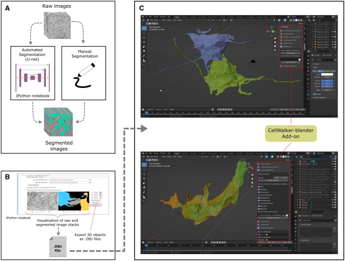
Summary: The implementation of computational tools for analysis of microscopy images has been one of the most important technological innovations in biology, providing researchers unmatched capabilities to comprehend cell shape and connectivity. While numerous tools exist for image annotation and segmentation, there is a noticeable gap when it comes to morphometric analysis of microscopy images. Most existing tools often measure features solely on 2D serial images, which can be difficult to extrapolate to 3D. For this reason, we introduce CellWalker, a computational toolbox that runs inside Blender, an open-source computer graphics software. This add-on improves the morphological analysis by seamlessly integrating analysis tools into the Blender workflow, providing visual feedback through a powerful 3D visualization, and leveraging the resources of Blender's community. CellWalker provides several morphometric analysis tools that can be used to calculate distances, volume, surface areas and to determine cross-sectional properties. It also includes tools to build skeletons, calculate distributions of subcellular organelles. In addition, this python-based tool contains 'visible-source' IPython notebooks accessories for segmentation of 2D/3D microscopy images using deep learning and visualization of the segmented images that are required as input to CellWalker. Overall, CellWalker provides practical tools for segmentation and morphological analysis of microscopy images in the form of an open-source and modular pipeline which allows a complete access to fine-tuning of algorithms through visible-source code while still retaining a result-oriented interface.
Availability and implementation: CellWalker source code is available on GitHub (https://github.com/utraf-pasteur-institute/Cellwalker-blender and https://github.com/utraf-pasteur-institute/Cellwalker-notebooks) under a GPL-3 license.
© The Author(s) 2023. Published by Oxford University Press.
Conflict of interest statement
None declared.
Figures
Similar articles
-
NeuroPycon: An open-source python toolbox for fast multi-modal and reproducible brain connectivity pipelines.Neuroimage. 2020 Oct 1;219:117020. doi: 10.1016/j.neuroimage.2020.117020. Epub 2020 Jun 6. Neuroimage. 2020. PMID: 32522662
-
Blik is an extensible 3D visualisation tool for the annotation and analysis of cryo-electron tomography data.PLoS Biol. 2024 Apr 30;22(4):e3002447. doi: 10.1371/journal.pbio.3002447. eCollection 2024 Apr. PLoS Biol. 2024. PMID: 38687779 Free PMC article.
-
STSE: Spatio-Temporal Simulation Environment Dedicated to Biology.BMC Bioinformatics. 2011 Apr 28;12:126. doi: 10.1186/1471-2105-12-126. BMC Bioinformatics. 2011. PMID: 21527030 Free PMC article.
-
Deep learning -- promises for 3D nuclear imaging: a guide for biologists.J Cell Sci. 2022 Apr 1;135(7):jcs258986. doi: 10.1242/jcs.258986. Epub 2022 Apr 14. J Cell Sci. 2022. PMID: 35420128 Free PMC article. Review.
-
Defining the boundaries: challenges and advances in identifying cells in microscopy images.Curr Opin Biotechnol. 2024 Feb;85:103055. doi: 10.1016/j.copbio.2023.103055. Epub 2023 Dec 23. Curr Opin Biotechnol. 2024. PMID: 38142646 Free PMC article. Review.
References
Publication types
MeSH terms
Grants and funding
LinkOut - more resources
Full Text Sources
Miscellaneous
