Gastrointestinal Kaposi Sarcoma without Dermatological Lesions: A Case Report
- PMID: 38060456
- PMCID: PMC10711638
- DOI: 10.12659/AJCR.941815
Gastrointestinal Kaposi Sarcoma without Dermatological Lesions: A Case Report
Abstract
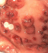
BACKGROUND Kaposi sarcoma is a malignancy of the vascular endothelium. It is associated with human herpesvirus 8 (HHV-8) infection, typically found with HIV/AIDS. It is rarely seen presenting as visceral involvement without any cutaneous lesions. Few case reports have described this. CASE REPORT We report a case of visceral Kaposi sarcoma (specifically, gastrointestinal lesions) without any cutaneous lesions in a 35-year-old man with HIV/AIDS who presented with abdominal pain, fatigue, and melena of a 15-day duration. Physical examination revealed tachycardia and hypertension, with a negative orthostatic sign. There were no visible signs of bleeding or cutaneous lesions, no abdominal pain, and a digital rectal examination was negative. Laboratory test results were significant for severe microcytic anemia, with hemoglobin level of 3.3 g/dL, decreased ferritin and iron levels, high red cell distribution width, and reticulocyte index lower than appropriate for anemia level. The absolute CD4 count was 33/uL, and the viral load was 56 895 copies/mL. Hemoglobin was optimized with packed red cells prior to endoscopy, and Pneumocystis jirovecii pneumonia prophylaxis was started. Esophagogastroduodenoscopy and colonoscopy revealed small and large bowel hemorrhagic stellate and annular lesions of varying sizes. Pathology reports from biopsy of the lesions seen in the procedure reported Kaposi sarcoma positive for HHV-8. He underwent chemotherapy with doxorubicin and showed clinical and laboratory improvement after treatment. CONCLUSIONS Kaposi sarcoma should be considered and investigated in patients with HIV/AIDS who are not on highly active antiretroviral therapy and present with gastrointestinal bleeding as an initial symptom, without any cutaneous lesions.
Conflict of interest statement
Figures


References
-
- Balachandra B, Tunitsky E, Dawood S, et al. Classic Kaposi’s sarcoma presenting first with gastrointestinal tract involvement in a HIV-negative Inuit male – a case report and review of the literature. Pathol Res Pract. 2006;202(8):623–26. - PubMed
-
- Kolios G, Kaloterakis A, Filiotou A, et al. Gastroscopic findings in Mediterranean Kaposi’s sarcoma (non-AIDS) Gastrointest Endosc. 1995;42(4):336–39. - PubMed
-
- Stebbing J, Sanitt A, Nelson M, et al. A prognostic index for AIDS-associated Kaposi’s sarcoma in the era of highly active antiretroviral therapy. Lancet. 2006;367(9521):1495–502. - PubMed
-
- Ahmed N, Nelson RS, Goldstein HM, Sinkovics JG. Kaposi’s sarcoma of the stomach and duodenum: Endoscopic and roentgenologic correlations. Gastrointest Endosc. 1975;21(4):149–52. - PubMed
Publication types
MeSH terms
Substances
LinkOut - more resources
Full Text Sources
Medical
Research Materials

