. 2023 Dec 7;9(1):161.
doi: 10.1038/s41531-023-00604-y.

Development and validation of an expanded antibody toolset that captures alpha-synuclein pathological diversity in Lewy body diseases

Affiliations

Development and validation of an expanded antibody toolset that captures alpha-synuclein pathological diversity in Lewy body diseases

Melek Firat Altay et al. NPJ Parkinsons Dis. .

Erratum in

Abstract

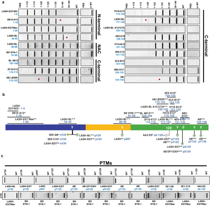
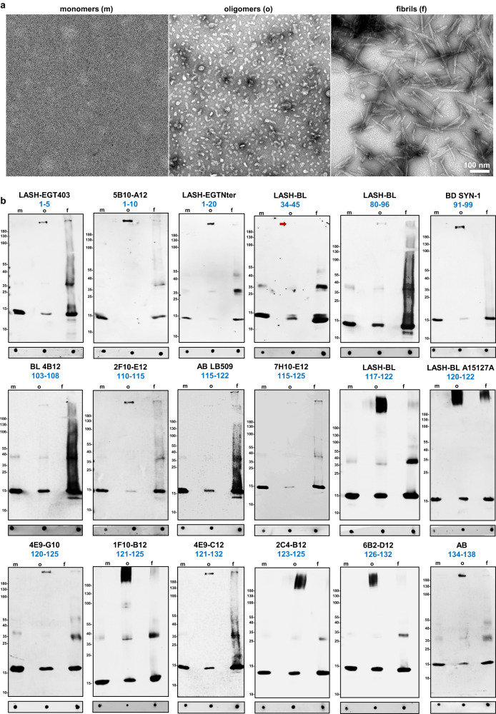
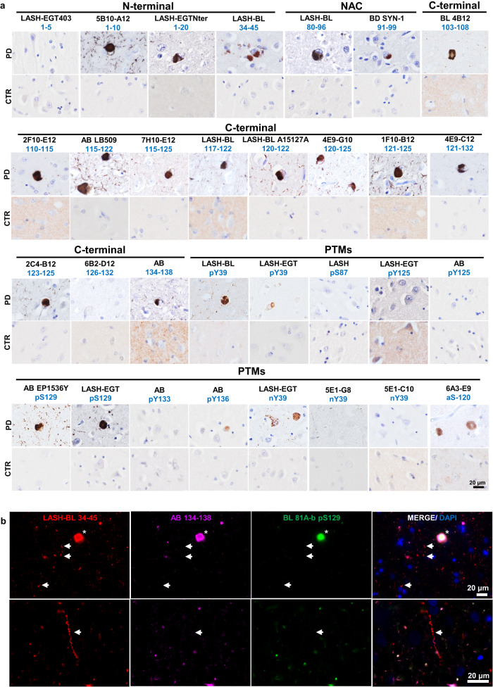
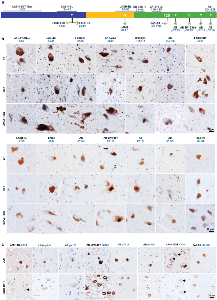
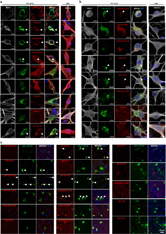
The abnormal aggregation and accumulation of alpha-synuclein (aSyn) in the brain is a defining hallmark of synucleinopathies. Various aSyn conformations and post-translationally modified forms accumulate in pathological inclusions and vary in abundance among these disorders. Relying on antibodies that have not been assessed for their ability to detect the diverse forms of aSyn may lead to inaccurate estimations of aSyn pathology in human brains or disease models. To address this challenge, we developed and characterized an expanded antibody panel that targets different sequences and post-translational modifications along the length of aSyn, and that recognizes all monomeric, oligomeric, and fibrillar aSyn conformations. Next, we profiled aSyn pathology across sporadic and familial Lewy body diseases (LBDs) and reveal heterogeneous forms of aSyn pathology, rich in Serine 129 phosphorylation, Tyrosine 39 nitration and N- and C-terminal tyrosine phosphorylations, scattered both to neurons and glia. In addition, we show that aSyn can become hyperphosphorylated during processes of aggregation and inclusion maturation in neuronal and animal models of aSyn seeding and spreading. The validation pipeline we describe for these antibodies paves the way for systematic investigations into aSyn pathological diversity in the human brain, peripheral tissues, as well as in cellular and animal models of synucleinopathies.


Conflict of interest statement

H.A.L. is the co-founder and chief scientific officer of ND BioSciences, Epalinges, Switzerland, a company that develops diagnostics and treatments for neurodegenerative diseases (NDs) based on platforms that reproduce the complexity and diversity of proteins implicated in NDs and their pathologies. H.A.L. has received funding from industry to support research on neurodegenerative diseases, including Merck Serono, UCB, and Abbvie. These companies had no specific role in the conceptualization, preparation, and decision to publish this manuscript. H.A.L. is the associate editor of npj Parkinson’s Disease. H.A.L. was not involved in the journal’s review of, or decisions related to, this manuscript. M.F.A., S.T.K., J.B., S.J., C.S., Y.M., L.P. and J.L.H. declare no competing interests.

Figures

Fig. 1
Fig. 1. Comprehensive exploration of aSyn heterogeneity - pathology diversity, fibril polymorphism, aSyn PTMs in synucleinopathy brains and antibody generation/ validation steps.
a Diversity of aSyn pathology in synucleinopathies with aa granular/punctate cytoplasmic inclusions in the neurons; ab classical LBs in the neuronal soma; ac LNs in the neuronal processes; ad astrocytic aSyn accumulations; ae oligodendroglial cytoplasmic inclusions. These pathological structures show differences in their positivity to aggregation markers, including ubiquitin (Ub) and p62. Schematic created with BioRender.com (agreement no: QW23G6FJ76). b Cryo-EM three-dimensional reconstructions of the recombinant full-length aSyn PFFs to show the polymorphism of aSyn fibrils generated in vitro,. Four distinct polymorphs were identified based on the protofilament fold and inter-protofilament interfaces: Polymorph 1a ‘rod’ (PDB-6CU7, EMD-7618); polymorph 1b ‘twister’ (PDB-6CU8, EMD-7619); polymorph 2a (PDB-6SSX, EMD-10307); and polymorph 2b (PDB-6SST, EMD-10305). c aSyn PTMs identified in synucleinopathy brain tissues, which include acetylation,,,, ubiquitination,,,,–, phosphorylation,,,–, nitration,, and truncation,,,,,,– across the whole sequence of the protein. d A schematic representation of the steps followed for the generation, characterization, validation and application of aSyn antibodies. These involved da antibody design via the selection of immunogens comprising of aSyn recombinant proteins and peptides; db immunization of the mice followed by lymphocyte–myeloma fusion; dc screening of the hybridomas via ELISA, DB and WB, isotyping and subcloning, and dd acquisition of purified antibodies. The antibodies were then de characterized using a library of aSyn recombinant proteins for their epitopes, conformational selectivity, sensitivity, specificity and reactivity via DB and WB. The antibody specificity was further validated on df aSyn KO mouse primary hippocampal and cortical neurons, and in aSyn KO mouse tissue of amygdala. dg The antibodies were validated on human brain tissues of different LB diseases. dh The mouse aSyn-reactive antibodies were applied to neuronal seeding model and PFF-injected mouse brain tissues to profile the newly formed aggregates. Schematic created with BioRender.com (agreement no: FN23G6E1SR). aSyn alpha-synuclein, DB dot/slot blot, cryo-EM cryogenic electron microscopy, ELISA enzyme-linked immunoassay, KO knockout, LB Lewy body, LN Lewy neurite, PFFs pre-formed fibrils, PTM post-translational modification, Ub ubiquitin, WB Western blot.
Fig. 2
Fig. 2. Validation and epitope mapping of aSyn antibodies.
a DB validation of the novel monoclonal, in-house polyclonal and commercially available aSyn antibodies against the N-terminal, NAC and the C-terminal regions of aSyn for epitope mapping, specificity and species reactivity using a selected library of aSyn recombinant proteins under native conditions. Protein loading control was run via Ponceau S staining. All loaded proteins represent human aSyn forms except for mouse aSyn wild-type (m WT) protein. Red arrows highlight the sensitivities of the antibodies to the presence of neighboring aSyn PTMs. b A schematic to represent the novel monoclonal (marked with *), in-house polyclonal (marked with **) and pre-existing commercial aSyn antibodies (marked with ***) included in this study. The commercial antibodies developed jointly with Biolegend are marked with *°*. The epitope information of each antibody is indicated in blue. Schematic created with BioRender.com (agreement no: JR23G6G5LA). c Specificity validation of the aSyn PTM antibodies via DB screening. aSyn alpha-synuclein, CTR control, DB dot/slot blot, FL full-length, m mouse, NAC non-amyloid component, PBS phosphate-buffered saline, PTM post-translational modification, WT wild-type.
Fig. 3
Fig. 3. Selectivity of aSyn antibodies over aSyn conformations.
a Representative EM images of aSyn human WT monomers, oligomers and fibrils. b DB and WB characterization of 18 aSyn antibodies to determine their conformational selectivity using aSyn human WT recombinant monomers, oligomers and fibrils. aSyn alpha-synuclein, DB dot/slot blot, EM electron microscopy, f fibrils, m monomers, o oligomers, WB Western blot, WT wild-type.
Fig. 4
Fig. 4. Application and validation of the aSyn antibodies on PD tissue.
a The in-house monoclonal, polyclonal and commercial aSyn antibodies were optimized for IHC on the PD cingulate cortex. Non-specific staining was not observed in age-matched healthy controls. Representative images taken from the cortical deep grey matter (layers V–VI) of PD1, CTR1 and CTR2. b Triple labeling of PD cingulate cortex by IF using an aSyn N-terminal (LASH-BL 34–45), a C-terminal (AB 134–138) and a pS129 (BL 81A-biotin) antibody. LBs are marked with asterisks, and LNs with arrows. Representative images from PD1 cingulate cortex are taken using Leica DM5500 B upright microscope at ×20 magnification. aSyn alpha-synuclein, CTR control, DAPI 4’,6-diamidino-2-phenylindole, IF immunofluorescence, IHC immunohistochemistry, LB Lewy body, LN Lewy neurite, NAC non-amyloid component, PD Parkinson’s disease, PTM post-translational modification.
Fig. 5
Fig. 5. A selected panel of aSyn antibodies reveal the broad diversity of human pathology in the SN of LBDs.
a An outline to show the epitopes of the aSyn antibody selection used for IHC studies on LBD tissues. Schematic created with BioRender.com (agreement no: NU23G6E7KK). b Representative images from the substantia nigra of sporadic (PD, DLB) and familial (SNCA H50Q) LBDs screened with the selection of aSyn non-modified and PTM antibodies. Images taken from the SN of PD2, PD3, DLB1, and SNCA H50Q1. c Representative images from the cingulate cortex of sporadic (DLB) and familial (SNCA G51D) LBDs screened with the selected aSyn PTM antibodies. Images taken from the cortical deep grey matter (layers V–VI) of DLB1 and SNCA G51D1. Arrows indicate star-shaped astroglial aSyn accumulations. aSyn alpha-synuclein, DLB dementia with Lewy bodies, IHC immunohistochemistry, LBD Lewy body disease, SN substantia nigra, PD Parkinson’s disease, PTM post-translational modification.
Fig. 6
Fig. 6. Application of the aSyn antibodies to the cellular and animal seeding models to profile the newly formed aSyn aggregates.
WT hippocampal neurons were seeded with PFFs for 14 days, and the newly formed aggregates monitored by ICC using the mouse-reactive a non-modified aSyn and b aSyn PTM antibodies in parallel to aSyn pS129 antibodies BL 81A or AB MJF-R13. c The same type of screening was run in PFF-injected mouse amygdala tissues by IHC. The non-modified aSyn antibody signals overlapping with the aSyn pS129-positive aggregates are marked with an arrow. The punctate positivity shown by aSyn pY39, pY133, and pY136 antibodies in close proximity to aSyn pS129-positive aggregates are shown by arrowheads. Note the non-specific diffuse positivity revealed by the two monoclonal nY39 antibodies 5E1-G8 and 5E1-C10 in the WT hippocampal neurons are also revealed in the aSyn KO neurons using these two antibodies (Supplementary Fig. 4A, B). aSyn alpha-synuclein, DAPI 4’,6-diamidino-2-phenylindole, ICC immunocytochemistry, IHC immunohistochemistry, KO knockout, MAP2 microtubule-associated protein 2, PBS phosphate-buffered saline, PFF pre-formed fibril, PTM post-translational modification, WT wild-type.

Similar articles

Cited by

References

    1. Baba M, et al. Aggregation of alpha-synuclein in Lewy bodies of sporadic Parkinson’s disease and dementia with Lewy bodies. Am. J. Pathol. 1998;152:879–884. - PMC - PubMed
    1. Spillantini, M. G. et al. Filamentous a-synuclein inclusions link multiple system atrophy with Parkinson’s disease and dementia with Lewy bodies. Neurosci. Lett.251, 205–208 (1998). - PubMed
    1. Spillantini MG, Crowther RA, Jakes R, Hasegawa M, Goedert M. Alpha-synuclein in filamentous inclusions of Lewy bodies from Parkinson’s disease and dementia with Lewy bodies. Proc. Natl. Acad. Sci. USA. 1998;95:6469–6473. doi: 10.1073/pnas.95.11.6469. - DOI - PMC - PubMed
    1. Appel-Cresswell S, et al. Alpha-synuclein p.H50Q, a novel pathogenic mutation for Parkinson’s disease: α- Synuclein p.H50q, a novel mutation For Pd. Mov. Disord. 2013;28:811–813. doi: 10.1002/mds.25421. - DOI - PubMed
    1. Chartier-Harlin M-C, et al. α-synuclein locus duplication as a cause of familial Parkinson’s disease. Lancet. 2004;364:1167–1169. doi: 10.1016/S0140-6736(04)17103-1. - DOI - PubMed