. 2023 Dec 7;14(1):8109.
doi: 10.1038/s41467-023-43906-8.

The amygdala is not necessary for the familiarity aspect of recognition memory

Affiliations

The amygdala is not necessary for the familiarity aspect of recognition memory

Benjamin M Basile et al. Nat Commun. .

Abstract

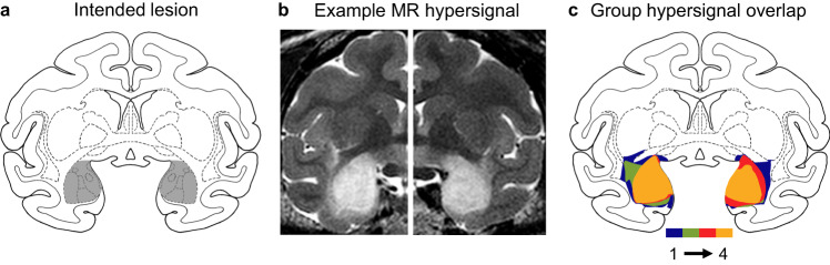
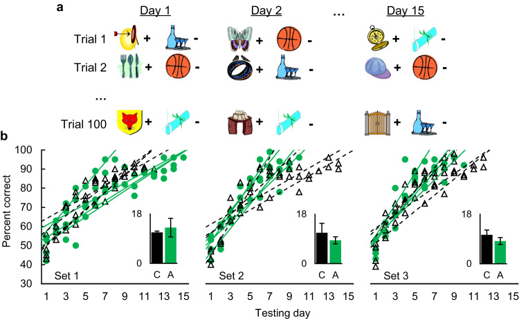
Dual-process accounts of item recognition posit two memory processes: slow but detailed recollection, and quick but vague familiarity. It has been proposed, based on prior rodent work, that the amygdala is critical for the familiarity aspect of item recognition. Here, we evaluated this proposal in male rhesus monkeys (Macaca mulatta) with selective bilateral excitotoxic amygdala damage. We used four established visual memory tests designed to assess different aspects of familiarity, all administered on touchscreen computers. Specifically, we assessed monkeys' tendencies to make low-latency false alarms, to make false alarms to recently seen lures, to produce curvilinear ROC curves, and to discriminate stimuli based on repetition across days. Three of the four tests showed no familiarity impairment and the fourth was explained by a deficit in reward processing. Consistent with this, amygdala damage did produce an anticipated deficit in reward processing in a three-arm-bandit gambling task, verifying the effectiveness of the lesions. Together, these results contradict prior rodent work and suggest that the amygdala is not critical for the familiarity aspect of item recognition.


Conflict of interest statement

The authors declare no competing interests.

Figures

Fig. 1
Fig. 1. Four rhesus monkeys received bilateral, selective, neurotoxic lesions of the amygdala.
a Diagram of a coronal section of the rhesus monkey brain at the level of the anterior commissure (+17 mm anterior to the auditory canal). The gray shaded region shows the location and extent of the intended lesion. b Example images from T2-weighted MR scans from one monkey in the amygdala lesion group acquired ~4 days after surgery in each hemisphere. The white hypersignal over the amygdala indicates edema consequent to injections of neurotoxins. c Overlap of observed hypersignal for the four monkeys in the lesion group. Colors indicate signal overlap from 1–4 monkeys.
Fig. 2
Fig. 2. Amygdala damage did not reduce the low-latency false alarms associated with reliance on quick familiarity.
a Diagram of the yes/no recognition test showing a match trial (top) and a nonmatch trial (bottom). Monkeys earned food by touching the test image if it matched the image remembered from study (a hit) or the nonmatch symbol if it did not (a correct rejection). b Error rates (±SEM) as a function of response speed decile (10% of responses/bin). Preoperative (top) and postoperative (bottom) performance is shown for control monkeys (n = 4; left) and monkeys with amygdala damage (n = 4; right). Postoperative data shown here are from the immediate postoperative test with the same stimuli. Closed symbols indicate false alarms (second-order polynomial fit with solid line) and open symbols indicate misses (linear fit with dashed line). Source data are provided as a Source data file.
Fig. 3
Fig. 3. Amygdala damage reduced false alarms to highly familiar probe lures.
a Example screens from the yes/no recognition task. Baseline match and nonmatch trials proceeded as in Exp 1. Probe trials were the same as nonmatch trials except that the to-be-rejected lure was the sample from the previous trial. b False alarm rates as a function of surgical timepoint (preoperative or postoperative on left and right, respectively, of each pair of bars), trial type (baseline or probe), group (control, n = 4, or amygdala, n = 4), and retention interval (4 or 20 s). Bars show group means and points show individual monkeys. * indicates p = 0.014 via two-sided, uncorrected, paired t-test. Source data are provided as a Source data file.
Fig. 4
Fig. 4. Amygdala damage did not change ROC measures of recollection or familiarity.
a We manipulated the decision criterion of the monkeys by changing the amount of food reward for hits and correct rejections each day. For example, five pellets for a correct rejection and one pellet for a hit should produce a very conservative bias to respond ‘no’ when uncertain. b The dual-process signal detection model posits that detailed recollection produces a flat but asymmetrical ROC curve, vague familiarity produces a curved but symmetrical ROC curve, and normal recognition produces a curved and asymmetrical ROC curve that represents the combination of recollection and familiarity. c The mean observed criterion bias (c’ ±SEM) of our monkeys as a result of the food manipulations in panel a. Negative numbers are more conservative biases and positive numbers are more liberal biases. These data indicate that the decision criteria of our monkeys were affected by the reward manipulations, as intended. Monkeys with amygdala damage (n = 4) in green circles and control monkeys in black triangles (n = 4). d The ROC curves for both control (left, black triangles, n = 4) and amygdala (right, green circles, n = 4) monkeys were curved and asymmetrical. Points represent group means of hit and false alarm rates at the five criterion bias levels. Inset bar graphs depict the mean (±SEM) of the individual monkeys’ parameter estimates for recollection (R) and familiarity (F). Source data are provided as a Source data file.
Fig. 5
Fig. 5. Amygdala damage did not impair across-day familiarity discrimination.
a Example stimuli used in each two-choice trial; + denotes a rewarded image and – denotes a nonrewarded image. The familiarity discrimination was such that novel images were always rewarded and familiar images were never rewarded (except for on Day 1, when all images were novel and reward assignments were initially unknown to the monkey). b Percent correct as a function of group (black triangles with dashed lines = control, n = 4; green circles with solid lines = amygdala, n = 4), testing day, and problem set. Each line is a linear fit for an individual monkey. Inset bar graphs depict the mean sessions to criterion (±SD) for each group (C = control, A = amygdala) and problem set. Source data are provided as a Source data file.
Fig. 6
Fig. 6. Amygdala damage impaired rapid reward association learning.
a Trial schematic. Examples screens (left) show the stimuli as they appeared to the monkey. Probabilities (right) show the pre-assigned reward probabilities of the images. The red question marks indicate a novel stimulus with an unknown (to the monkey) probability that must be learned. b Model-derived learning parameters for control (black bars, n = 4) and amygdala (green bars, n = 4) monkeys. Learning rate (α) quantifies how much learning occurs from each outcome; Inverse temperature (β) quantifies the consistency with which subjects select the most valuable option; Novelty bonus quantifies the bias to choose or avoid a novel item with an unknown reward probability. Bars are group means and points are parameters (±95% CI) for individual monkeys. * indicates p = 0.035 via linear mixed-effects model as described in text. c Proportion (±SEM) of times a novel option was chosen as a function of assigned reward probability (75%, 50%, or 25%) and number of trials since the introduction of a novel item. Trial 0 is the trial on which the novel item was introduced. Horizontal lines below the data indicate trials for which two conditions were chosen at statistically different rates (uncorrected = thin line; Bonferroni corrected = thick line; HvL = high reward vs. low reward; MvL = medium reward vs. low reward; MvH = medium reward vs. high reward). Vertical lines indicate the trial on which all three conditions were chosen at statistically different levels (uncorrected). Compare panel c to Fig. 1E from Costa et al. (2019). Source data are provided as a Source data file.

Similar articles

Cited by

References

    1. Yonelinas AP. The nature of recollection and familiarity: a review of 30 years of research. J. Mem. Lang. 2002;46:441–517. doi: 10.1006/jmla.2002.2864. - DOI
    1. Brainerd CJ, Nakamura K, Lee W-F. Recollection is fast and slow. J. Exp. Psychol. Learn. Mem. Cogn. 2019;45:302. doi: 10.1037/xlm0000588. - DOI - PMC - PubMed
    1. Ingram KM, Mickes L, Wixted JT. Recollection can be weak and familiarity can be strong. J. Exp. Psychol. Learn Mem. Cogn. 2012;38:325–339. doi: 10.1037/a0025483. - DOI - PMC - PubMed
    1. Brandt KR, Gardiner JM, Vargha-Khadem F, Baddeley AD, Mishkin M. Impairment of recollection but not familiarity in a case of developmental amnesia. Neurocase. 2009;15:60–65. doi: 10.1080/13554790802613025. - DOI - PMC - PubMed
    1. Mayes AR, Holdstock JS, Isaac CL, Hunkin NM, Roberts N. Relative sparing of item recognition memory in a patient with adult-onset damage limited to the hippocampus. Hippocampus. 2002;12:325–340. doi: 10.1002/hipo.1111. - DOI - PubMed

Publication types