. 2023 Dec 7;13(1):21618.
doi: 10.1038/s41598-023-48072-x.

Cortico-muscular connectivity is modulated by passive and active Lokomat-assisted Gait

Affiliations

Cortico-muscular connectivity is modulated by passive and active Lokomat-assisted Gait

Fiorenzo Artoni et al. Sci Rep. .

Abstract

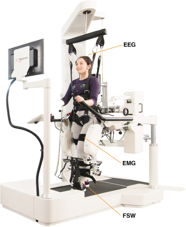
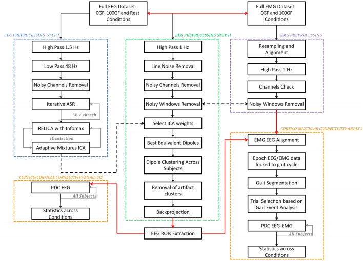
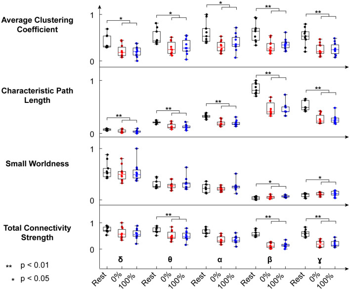
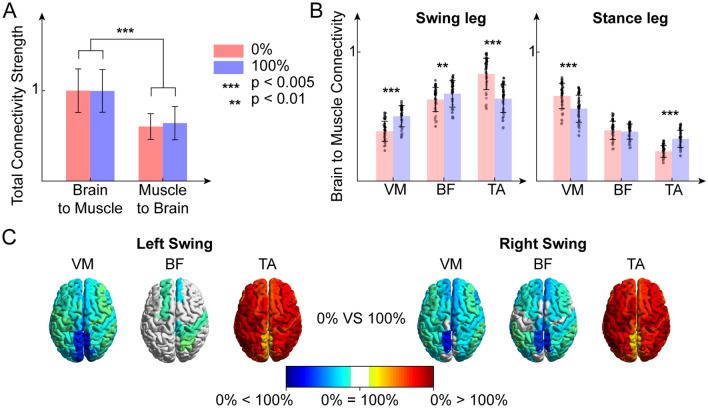
The effects of robotic-assisted gait (RAG) training, besides conventional therapy, on neuroplasticity mechanisms and cortical integration in locomotion are still uncertain. To advance our knowledge on the matter, we determined the involvement of motor cortical areas in the control of muscle activity in healthy subjects, during RAG with Lokomat, both with maximal guidance force (100 GF-passive RAG) and without guidance force (0 GF-active RAG) as customary in rehabilitation treatments. We applied a novel cortico-muscular connectivity estimation procedure, based on Partial Directed Coherence, to jointly study source localized EEG and EMG activity during rest (standing) and active/passive RAG. We found greater cortico-cortical connectivity, with higher path length and tendency toward segregation during rest than in both RAG conditions, for all frequency bands except for delta. We also found higher cortico-muscular connectivity in distal muscles during swing (0 GF), and stance (100 GF), highlighting the importance of direct supraspinal control to maintain balance, even when gait is supported by a robotic exoskeleton. Source-localized connectivity shows that this control is driven mainly by the parietal and frontal lobes. The involvement of many cortical areas also in passive RAG (100 GF) justifies the use of the 100 GF RAG training for neurorehabilitation, with the aim of enhancing cortical-muscle connections and driving neural plasticity in neurological patients.


Conflict of interest statement

The authors declare no competing interests.

Figures

Figure 1
Figure 1
Lokomat exoskeleton. Figure representing the robotic device used. In this study, subjects are supported by a harness providing up to full body weight support while performing gait training. The exoskeleton was used both actively (0% guidance force support) and passively (100% guidance force support). While using the exoskeleton, 4 footswitches (FSW), 6 bipolar wireless EMG electrodes and a 64 channels EEG cap were allowed to image the brain/body activity during the gait tasks. Figure adapted from the website https://www.hocoma.com.
Figure 2
Figure 2
Full process pipeline. The EEG data underwent two preprocessing steps, namely EEG Preprocessing Step I (blue box) and EEG Preprocessing Step II (green box). Independent Components (ICs) were extracted within Step I. IC weights were then transferred to the dataset, conservatively processed according to Step II. Step I is designed to extract ICs with the best possible reliability. Step II is more conservative, and it is designed to retain the maximum amount of information for subsequent connectivity analyses. Each IC was source-localized using a dipolar model, and artifact clusters of ICs were removed from the data. The backprojected EEG data was then used to perform distributed source localization, and the AAL atlas EEG ROIs time courses were extracted. Cortico-cortical connectivity analysis was then performed across all subjects for 0 GF, 100 GF, and Rest conditions using the time course of the source-localized activity within the regions of interest (ROIs) extracted after Step II. In parallel, EMG data were aligned to EEG, high-passed at 2 Hz and noisy windows labeled for rejection. Only time windows that were not labeled as artifact either for EEG or EMG were retained. Cortico-muscular connectivity (assessed through Partial Directed Coherence—PDC) was estimated for 0 GF and 100 GF respectively on epoched data, time locked to the gait cycle (see “Methods” for more details). Group-level statistical analysis across all subjects and conditions was then performed.
Figure 3
Figure 3
Normalized network features (average path length, small worldness, clustering coefficient and total connectivity strength) of the intracortical connectivity network in the 3 conditions: rest (black), 0% GF (blue) and 100% GF (red).
Figure 4
Figure 4
(A) Brain-to-muscle normalized connectivity strength and muscle-to-brain normalized connectivity strength for both guidance force levels. (B) Normalized brain to muscle connectivity flow toward vastus medialis (VM), tibialis anterior (TA) and biceps femoris (BF) of the swing leg (left) and of the stance leg (right), for both guidance force levels. (C) Brain maps of the significative difference in cortico-muscular connectivity between guidance force levels, divided by muscle.
Figure 5
Figure 5
Bar plots of the cortico-muscular connections values directed from the parietal lobes (pink), frontal lobes (yellow), and the posterior fossa (green) to the vastus medialis (VM), tibialis anterior (TA) and biceps femoris (BF) of the swing leg (left) and of the stance leg (right), for both guidance force levels (up: 0% GF, bottom: 100% GF).
Figure 6
Figure 6
(A) Normalized ROI outflow (sum of outcoming connections) in cortico-cortical connectivity in both guidance force levels (left) and normalized ROI outflow in cortico-muscular connectivity in both guidance force levels (right). (B) Normalized outflow of the ROIs of the posterior fossa in cortico-cortical connectivity and outflow of all other ROIs, for both guidance force levels. (C) Normalized outflow of the ROIs of the posterior fossa in cortico-muscular connectivity and outflow of all other ROIs, for both guidance force levels.

References

    1. Gimigliano F, et al. Robot-assisted arm therapy in neurological health conditions: Rationale and methodology for the evidence synthesis in the CICERONE Italian Consensus Conference. Eur. J. Phys. Rehabil. Med. 2021;57(5):824–830. doi: 10.23736/S1973-9087.21.07011-8. - DOI - PubMed
    1. Calabrò RS, et al. What does evidence tell us about the use of gait robotic devices in patients with multiple sclerosis? A comprehensive systematic review on functional outcomes and clinical recommendations. Eur. J. Phys. Rehabil. Med. 2021;7:841–849. - PubMed
    1. Carmignano SM, et al. Robot-assisted gait training in patients with Parkinson’s disease: Implications for clinical practice. A systematic review. NeuroRehabilitation. 2022;51:649–663. doi: 10.3233/NRE-220026. - DOI - PubMed
    1. Mehrholz J, Hädrich A, Platz T, Kugler J, Pohl M. Electromechanical and robot-assisted arm training for improving generic activities of daily living, arm function, and arm muscle strength after stroke. Cochrane Database Syst. Rev. 2012;6:CD006876. - PubMed
    1. Kleim, J. A. & Jones, T. A. Principles of experience-dependent neural plasticity: implications for rehabilitation after brain damage (2008). - PubMed