. 2023 Dec 8;14(12):804.
doi: 10.1038/s41419-023-06336-4.

Cancer CD39 drives metabolic adaption and mal-differentiation of CD4+ T cells in patients with non-small-cell lung cancer

Affiliations

Cancer CD39 drives metabolic adaption and mal-differentiation of CD4+ T cells in patients with non-small-cell lung cancer

Ying Wang et al. Cell Death Dis. .

Abstract

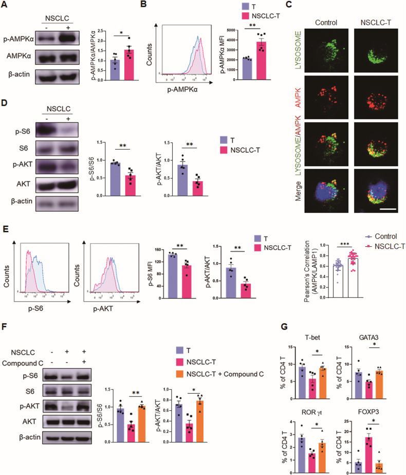
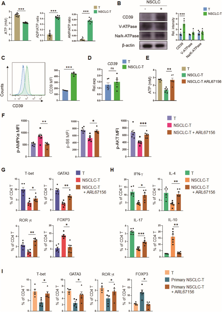
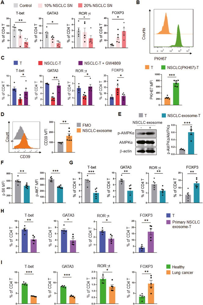
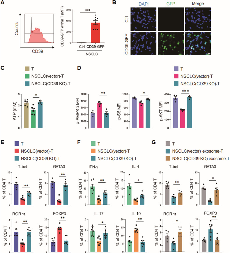
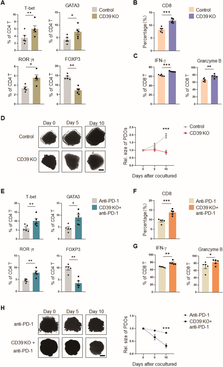
While ectonucleotidase CD39 is a cancer therapeutic target in clinical trials, its direct effect on T-cell differentiation in human non-small-cell lung cancer (NSCLC) remains unclear. Herein, we demonstrate that human NSCLC cells, including tumor cell lines and primary tumor cells from clinical patients, efficiently drive the metabolic adaption of human CD4+ T cells, instructing differentiation of regulatory T cells while inhibiting effector T cells. Of importance, NSCLC-induced T-cell mal-differentiation primarily depends on cancer CD39, as this can be fundamentally blocked by genetic depletion of CD39 in NSCLC. Mechanistically, NSCLC cells package CD39 into their exosomes and transfer such CD39-containing exosomes into interacting T cells, resulting in ATP insufficiency and AMPK hyperactivation. Such CD39-dependent NSCLC-T cell interaction holds well in patients-derived primary tumor cells and patient-derived organoids (PDOs). Accordingly, genetic depletion of CD39 alone or in combination with the anti-PD-1 immunotherapy efficiently rescues effector T cell differentiation, instigates anti-tumor T cell immunity, and inhibits tumor growth of PDOs. Together, targeting cancer CD39 can correct the mal-differentiation of CD4+ T cells in human NSCLC, providing in-depth insight into therapeutic CD39 inhibitors.


Conflict of interest statement

The authors declare no competing interests.

Figures

Fig. 1
Fig. 1. NSCLC drives mal-differentiation of CD4+ T cells.
AD CD4+ T cells isolated from PBMCs of healthy donors were pre-conditioned with human lung adenocarcinoma A549 cells for 12 hours, followed by anti-CD3/CD28 activation for 4 days, and detections of linage-determining transcription factors (A, B) and signature cytokines (C, D). E, F Healthy CD4+ T cells were pre-conditioned with NSCLC patients-derived primary tumor cells for 12 hours, followed by anti-CD3/CD28 activation for 4 days, and detection of linage-determining transcription factors. Mean ± SEM from 6 individuals in each group. *p < 0.05, **p < 0.01, ***p < 0.001 with paired student t-test.
Fig. 2
Fig. 2. NSCLC promotes AMPK activation for T cell mal-differentiation.
A, B Healthy CD4+ T cells were pre-conditioned with A549 cells for 12 h, activated with anti-CD3/CD28 beads for 3 days, and tested for p-AMPKα and AMPKα by Western blot and flow cytometry. Mean ± SEM from 5 individuals in each group. C Co-localization of AMPK with LAMP1+ lysosomes detected by confocal microscopy. A representative from 3 individuals per group. Scale bar, 5 μm. Mean ± SEM from 30 individuals in each group. D, E Healthy CD4+ T cells were pre-conditioned with A549 cells for 12 h, activated with anti-CD3/CD28 beads for 3 days, and detected for p-S6 and p-AKT. Mean ± SEM from 5 individuals in each group. F, G A549 cell pre-conditioned CD4+ T cells were activated with anti-CD3/CD28 beads for 3 days in the presence or absence of AMPK inhibitor Compound C (10 μM), followed by detection of T cell differentiation. Mean ± SEM from 5 individuals in each group. *p < 0.05, **p < 0.01 with paired student t-test (AE) and ANOVA plus Tukey method (F, G).
Fig. 3
Fig. 3. CD39 is crucial for NSCLC-instructed T cell mal-differentiation.
A Healthy CD4+ T cells were pre-conditioned with A549 cells for 12 h, activated with anti-CD3/CD28 beads for 3 days, and detected for energy status. Mean ± SEM from 8 individuals in each group. B Healthy CD4+ T cells were incubated with A549 cells for 12 h and detected for the expressions of CD39, V-ATPase, and Na/K ATPase by western blot. Mean ± SEM from 4 individuals in each group. C Healthy CD4+ T cells were incubated with A549 cells for 12 h and detected for CD39 protein levels by flow cytometry. Mean ± SEM from 5 individuals in each group. D Healthy CD4+ T cells were incubated with A549 cells for 12 h and detected for CD39 mRNA expressions. Mean ± SEM from 6 individuals in each group. E, F Healthy CD4+ T cells were pre-conditioned with A549 cells in the presence or absence of CD39 inhibitor ARL67156 (10 μM) for 12 hours and activated with anti-CD3/CD28 beads for 3 days. Mean ± SEM from 6 individuals in each group. G, H Healthy CD4+ T cells were pre-conditioned with A549 cells in the presence or absence of CD39 inhibitor ARL67156 (10 μM) for 12 h, followed by activation and detection of T cell differentiation. Mean ± SEM from 6 individuals in each group. I Healthy CD4+ T cells were pre-conditioned with NSCLC patients-derived primary tumor cells in the presence or absence of CD39 inhibitor ARL67156 (10 μM) for 12 h, followed by activation and detection of T cell differentiation. Mean ± SEM from 5 individuals in each group. *p < 0.05, **p < 0.01, ***p < 0.001 with paired student t-test (AD) and ANOVA plus Tukey method (EI).
Fig. 4
Fig. 4. NSCLC transfer CD39+ exosomes for instructing T cell mal-differentiation.
A Healthy CD4+ T cells were pre-conditioned with conditioned media containing 10% and 20% A549 supernatant for 12 h, followed by activation and detection for T cell differentiation. Mean ± SEM from 5 individuals in each group. B Healthy CD4+ T cells were co-cultured with PKH67-labeled A549 cells for 12 h and detected for PKH67-labeled membrane vesicles. Mean ± SEM from 6 individuals in each group. C A549 cells were pre-treated with exosome inhibitor GW4869 (10 μM) for 24 h before pre-conditioning healthy CD4+ T cells for another 12 h. After that, CD4+ T cells were activated with anti-CD3/CD28 beads for 4 days and analyzed for T cell differentiation. Mean ± SEM from 5 individuals in each group. D CD39 protein in A549 cell-derived exosomes was detected by flow cytometry. Mean ± SEM from 6 individuals in each group. EG Healthy CD4+ T cells were pre-treated with A549-derived exosomes for 12 h and activated with anti-CD3/CD28 beads. AMPK activation and mTOR activity were detected 3 days post-stimulation, while T-cell differentiation was determined on day 4. Mean ± SEM from 5–6 individuals in each group. H Healthy CD4+ T cells were pre-conditioned with exosomes from NSCLC patient-derived primary tumor cells for 12 h and activated with anti-CD3/CD28 beads for 4 days, and detected for T cell differentiation by flow cytometry. Mean ± SEM from 5 individuals in each group. I CD4+ T cells isolated from healthy donors, or NSCLC patients, were activated with anti-CD3/CD28 beads for 4 days, and detected for T cell differentiation by flow cytometry. Mean ± SEM from 5 individuals in each group. *p < 0.05, **p < 0.01, ***p < 0.001 with ANOVA plus Tukey method (A, C), paired student t-test (B, DH) and unpaired student t-test (I).
Fig. 5
Fig. 5. Genetic knockout of CD39 in NSCLC abrogates NSCLC-T cell interaction.
A, B A549 cells were transfected with GFP-labeled CD39 expression vector, followed by incubation with healthy CD4+ T cells for 24 h. GFP-labeled CD39 protein in interacting T cells was determined with flow cytometry and confocal microscopy. Representative and mean ± SEM from 8 independent experiments. Scale bar 5 μm. C, D Healthy CD4+ T cells of healthy donors were pre-conditioned with CD39-deficient or control A549 cells for 12 h, followed by activation and detections for metabolic adaption. Mean ± SEM from 4–6 individuals in each group. E, F Healthy CD4+ T cells were pre-conditioned with CD39-deficient or control A549 cells for 12 h, followed by activation and analyses of T cell differentiation. Mean ± SEM from 6 individuals in each group. G Healthy CD4+ T cells were pre-conditioned with exosomes derived from CD39-deficient or control A549 cells for 12 h, followed by activation and analyses of T cell differentiation. Mean ± SEM from 6 individuals in each group. *p < 0.05, **p < 0.01, ***p < 0.001 with paired student t-test (A) and ANOVA plus Tukey method (CG).
Fig. 6
Fig. 6. Targeting CD39 restores effector T cell differentiation and instigates anti-tumor T cell immunity.
A PBMCs of NSCLC patients were co-cultured with corresponding PDOs or CD39 KO-PDOs for 10 days and detected for T cell differentiation. Mean ± SEM from 5 individuals in each group. B, C CD8+ T cells within the indicated PDOs were tested for productions of IFN-γ and Granzyme B by flow cytometry. Mean ± SEM from 5 individuals in each group. D CD39-deficient and control PDOs were incubated with the same patients’ PBMCs for the indicated time and analyzed for tumor outgrowth. Mean ± SEM from 5 individuals in each group. Scale bars, 200 μm. E CD39-deficient and control PDOs receiving anti-PD-1 antibody (10 μg/mL) treatment were incubated with the same patients’ PBMCs for 10 days and detected for T cell differentiation. Mean ± SEM from 5 individuals in each group. F, G PDO-infiltrating CD8+ T cells were analyzed for production of IFN-γ or Granzyme B by flow cytometry. Mean ± SEM from 5 individuals in each group. H CD39-deficient and control PDOs receiving anti-PD-1 antibody (10 μg/mL) treatment were incubated with the same patients’ PBMCs and analyzed for tumor growth at the indicated time. Mean ± SEM from 5 individuals in each group. Scale bars, 200 μm. *p < 0.05, **p < 0.01, ***p < 0.001 with paired student t-test.

Similar articles

Cited by

References

    1. J. F, M. E, F. L, M. C, L. M, Piñeros M ea. Global Cancer Observatory: Cancer Today. Lyon: International Agency for Research on Cancer; 2020. 2021 [cited 2021 August] Available from: https://gco.iarc.fr/today
    1. Reck M, Rodriguez-Abreu D, Robinson AG, Hui R, Csoszi T, Fulop A, et al. Pembrolizumab versus chemotherapy for PD-L1-positive non-small-cell lung cancer. N Engl J Med. 2016;375:1823–33. doi: 10.1056/NEJMoa1606774. - DOI - PubMed
    1. Socinski MA, Jotte RM, Cappuzzo F, Orlandi F, Stroyakovskiy D, Nogami N, et al. Atezolizumab for first-line treatment of metastatic nonsquamous NSCLC. N Engl J Med. 2018;378:2288–301. doi: 10.1056/NEJMoa1716948. - DOI - PubMed
    1. Perets R, Bar J, Rasco DW, Ahn MJ, Yoh K, Kim DW, et al. Safety and efficacy of quavonlimab, a novel anti-CTLA-4 antibody (MK-1308), in combination with pembrolizumab in first-line advanced non-small-cell lung cancer. Ann Oncol. 2021;32:395–403. doi: 10.1016/j.annonc.2020.11.020. - DOI - PubMed
    1. Schoenfeld JD, Giobbie-Hurder A, Ranasinghe S, Kao KZ, Lako A, Tsuji J, et al. Durvalumab plus tremelimumab alone or in combination with low-dose or hypofractionated radiotherapy in metastatic non-small-cell lung cancer refractory to previous PD(L)-1 therapy: an open-label, multicentre, randomised, phase 2 trial. Lancet Oncol. 2022;23:279–91. doi: 10.1016/S1470-2045(21)00658-6. - DOI - PMC - PubMed

Publication types