. 2023 Dec 7;13(1):21637.
doi: 10.1038/s41598-023-48365-1.

Platelet-activating factor and protease-activated receptor 2 cooperate to promote neutrophil recruitment and lung inflammation through nuclear factor-kappa B transactivation

Affiliations

Platelet-activating factor and protease-activated receptor 2 cooperate to promote neutrophil recruitment and lung inflammation through nuclear factor-kappa B transactivation

Irismara Sousa Silva et al. Sci Rep. .

Abstract

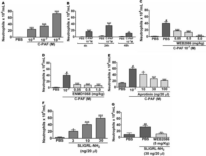
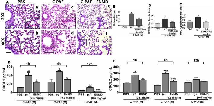
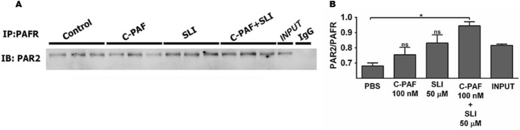
Although it is well established that platelet-activated receptor (PAF) and protease-activated receptor 2 (PAR2) play a pivotal role in the pathophysiology of lung and airway inflammatory diseases, a role for a PAR2-PAFR cooperation in lung inflammation has not been investigated. Here, we investigated the role of PAR2 in PAF-induced lung inflammation and neutrophil recruitment in lungs of BALB/c mice. Mice were pretreated with the PAR2 antagonist ENMD1068, PAF receptor (PAFR) antagonist WEB2086, or aprotinin prior to intranasal instillation of carbamyl-PAF (C-PAF) or the PAR2 agonist peptide SLIGRL-NH2 (PAR2-AP). Leukocyte infiltration in bronchoalveolar lavage fluid (BALF), C-X-C motif ligand 1 (CXCL)1 and CXCL2 chemokines, myeloperoxidase (MPO), and N-acetyl-glycosaminidase (NAG) levels in BALF, or lung inflammation were evaluated. Intracellular calcium signaling, PAFR/PAR2 physical interaction, and the expression of PAR2 and nuclear factor-kappa B (NF-КB, p65) transcription factor were investigated in RAW 264.7 cells stimulated with C-PAF in the presence or absence of ENMD1068. C-PAF- or PAR2-AP-induced neutrophil recruitment into lungs was inhibited in mice pretreated with ENMD1068 and aprotinin or WEB2086, respectively. PAR2 blockade impaired C-PAF-induced neutrophil rolling and adhesion, lung inflammation, and production of MPO, NAG, CXCL1, and CXCL2 production in lungs of mice. PAFR activation reduced PAR2 expression and physical interaction of PAR2 and PAFR; co-activation is required for PAFR/PAR2 physical interaction. PAR2 blockade impaired C-PAF-induced calcium signal and NF-κB p65 translocation in RAW 264.7 murine macrophages. This study provides the first evidence for a cooperation between PAFR and PAR2 mediating neutrophil recruitment, lung inflammation, and macrophage activation.


Conflict of interest statement

The authors declare no competing interests.

Figures

Figure 1
Figure 1
PAR2 blockade and inhibition of proteases impair PAF-induced neutrophil migration in BALB/c mice. (A,B) Mice were instilled intranasally with PBS or C-PAF. The number of infiltrating neutrophils was counted in BALF 4, 24, or 48 h after challenge. (CG) Mice were pretreated with an intraperitoneal injection of WEB 2086 or ENMD1068, or with intranasal instillation of PBS or aprotinin 1 h before intranasal instillation of C-PAF or SLIGRL-NH2. The number of infiltrating neutrophils was counted in BALF 24 h after challenge. The results are representative of three independent experiments and are expressed as mean ± SEM from six mice per group. Statistical analysis was assessed by ANOVA followed by Newman-Keuls test, A, B and F: ***p < 0.001 and *p < 0.05 compared to control group (PBS); C, D, E and G: ***p < 0.001, **p < 0.01 and *p < 0.05 compared to C-PAF group; C, D, E and F: #p < 0.001 or ##p < 0.01 compared to control group (PBS).
Figure 2
Figure 2
PAR2 blockade impairs rolling and adhesion of leukocytes in the mesentery microcirculation of mice. (AF) Representative imaging of mesentery microcirculation by intravital microscopy (neutrophils in red, anti-Ly6G/Ly6C-Gr-1; 0.2 mg/mL, i.v.). Mice were pretreated with ENMD1068 (0.5 mg/kg, i.p.) 1 h before stimulation with C-PAF (10−7 M, i.p.). After 4 h, rolling (G), velocity (H), and adhesion (I) of leukocytes (white arrows) were evaluated in the venules of mesentery microcirculation. The fluorescence intensity was monitored for 10 min using an objective Plan Apo 20 × calibrated by each venule. The results are representative of three independent experiments with similar results and are expressed as mean ± SEM from six mice per group. Statistical analysis was assessed by ANOVA followed by Newman-Keuls test, (GI) ***p < 0.001 compared to C-PAF group and #p < 0.001 compared to control group (PBS). Scale bar: 90 μm or 43 μm.
Figure 3
Figure 3
PAR2 blockade reduces C-PAF-induced lung inflammation and production of chemokines in BALB/c mice. (A) Mice were pretreated with ENMD1068 (0.5 mg/kg, i.p.). One hour later the mice were stimulated with C-PAF (10–7 M; i.n.). After 24 h, the lungs were collected and stained with H&E (20 × scale of 100 μM; 40 × scale of 50 μM). Panel a-b: PBS group, which exhibits normal histological features and no evidence of lung inflammation; Panel c-d: C-PAF group showing perivascular and peribronchiolar inflammation; Panel e–g: ENMD1068 pretreated group, shows cellular infiltration reduced as assessed by the histopathology score. (BE) mice were pretreated with ENMD1068 (0.5 mg/kg, i.p.) 1 h prior to administration of C-PAF (10–7 M; i.n.). NAG and MPO levels were evaluated 24 h later and CXCL1 and CXCL2 levels were assessed 1, 4, and 12 h later. The results are representative of three independent experiments and expressed as mean ± SEM from six mice per group. Statistical analysis was assessed by ANOVA followed by Newman-Keuls test, Panel g: ***p < 0.001 compared to C-PAF group and #p < 0.001 compared to control group (PBS); (B–E) ***p < 0.001, **p < 0.01 and *p < 0.05 compared to C-PAF group and #p < 0.001, ##p < 0.01 and ###p < 0.05 compared to control group (PBS).
Figure 4
Figure 4
PAFR/PAR2 physical interaction in RAW 264.7 murine macrophages. Cells were stimulated with C-PAF (100 nM), SLIGRL-NH2 (50 μM), or simultaneous co-stimulation (C-PAF 100 nM + SLIGRL-NH2 50 μM) for 20 min before addition of RIPA. Cell lysates were subjected to immunoprecipitation (A) and west immunoblotting (B) using antibodies to PAFR and PAR2. Representative image of the interaction and quantification of PAR2 and PAFR proteins immunoprecipitated. The experiments were performed in duplicate and one membrane was developed with anti-PAR2 and the other with anti-PAFR for normalization control. In data shown, each band in representative membrane image of western blotting correspond of three separate experiments realized before gel electrophoresis running. Values are expressed as mean ± SEM. Statistical analysis was assessed by ANOVA followed by Newman-Keuls test, B: *p < 0.05 compared to control group (PBS).
Figure 5
Figure 5
PAR2 blockade reduced C-PAF-induced intracellular Ca2+ signals in RAW 264 cells. Cells were pretreated with ENMD1068 (5 μM) 1 h before C-PAF (100 nM) stimulation. (AC) Representative cells for Ca2+ signal amplitude as a function of time. (D) Ca2+ signaling amplitude (% above basal) and (E) percentage of responsive cells (% above basal). Bars represent mean ± SEM (n = 3 preparations with 30 cells per group). Statistical analysis was by ANOVA followed by Newman-Keuls test, (C) and (E) ***p < 0.001 and *p < 0.05 compared to C-PAF group.
Figure 6
Figure 6
Expression of PAR2 and NF-КB (p65) transcription factor in RAW 264.7 cells. Cells were pretreated with ENMD1068 (5 μM) 1 h prior to C-PAF (100 nM) stimulation, fluorescence intensity was evaluated 4 h later. (A) Representative images of PAR2 (red), NF-kB (p65, green) and DAPI (nucleus, blue) expression. (B) PAR2 fluorescence intensity. (C) Nuclear fluorescence intensity for NF-κB (p65). (D) Cells were pretreated with ENMD1068 (5 μM) 1 h prior C-PAF (100 nM) stimulation. PAR2 mRNA was analyzed 1, 2, and 3 h later by qPCR. (B,C) Bars represent mean ± SEM (n = 3 preparations with 30 cells per group). Statistical analysis was by ANOVA followed by Newman-Keuls test, (B) and (D) ***p < 0.001 and *p < 0.05 compared to control group (PBS); (C) and (D) ***p < 0.001 and *p < 0.05 compared to C-PAF group and #p < 0.001 compared to control group (PBS).

References

    1. Moldoveanu B, et al. Inflammatory mechanisms in the lung. J. Inflam. Res. 2009;2:1–11. doi: 10.2147/JIR.S4385. - DOI - PMC - PubMed
    1. Aghasafari P, George U, Pidaparti R. A review of inflammatory mechanism in airway diseases. Inflam. Res. 2019;68(1):59–74. doi: 10.1007/s00011-018-1191-2. - DOI - PubMed
    1. Muñoz-Cano RM, et al. Platelet-activating factor (PAF) in allergic rhinitis: Clinical and therapeutic implications. J. Clin. Med. 2019;8(9):1338–1350. doi: 10.3390/jcm8091338. - DOI - PMC - PubMed
    1. Benveniste J, Henson PM, Cochrane CG. Leukocyte-dependent histamine release from rabbit platelets: The role of IgE, basophils, and a platelet-activating factor. J. Exper. Med. 1972;136(6):1356–1377. doi: 10.1084/jem.136.6.1356. - DOI - PMC - PubMed
    1. Benveniste J. Platelet-activating factor, a new mediator of anaphylaxis and immune complex deposition from rabbit and human basophils. Nature. 1974;249(457):581–582. doi: 10.1038/249581a0. - DOI - PubMed