

Management of Gastric Neuroendocrine Tumors: A Review
- PMID: 38062290
- PMCID: PMC10922891
- DOI: 10.1245/s10434-023-14712-9
Management of Gastric Neuroendocrine Tumors: A Review
Abstract
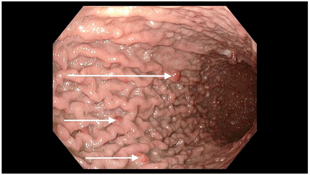
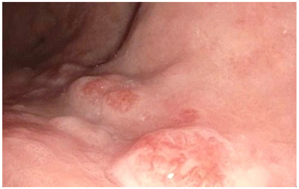
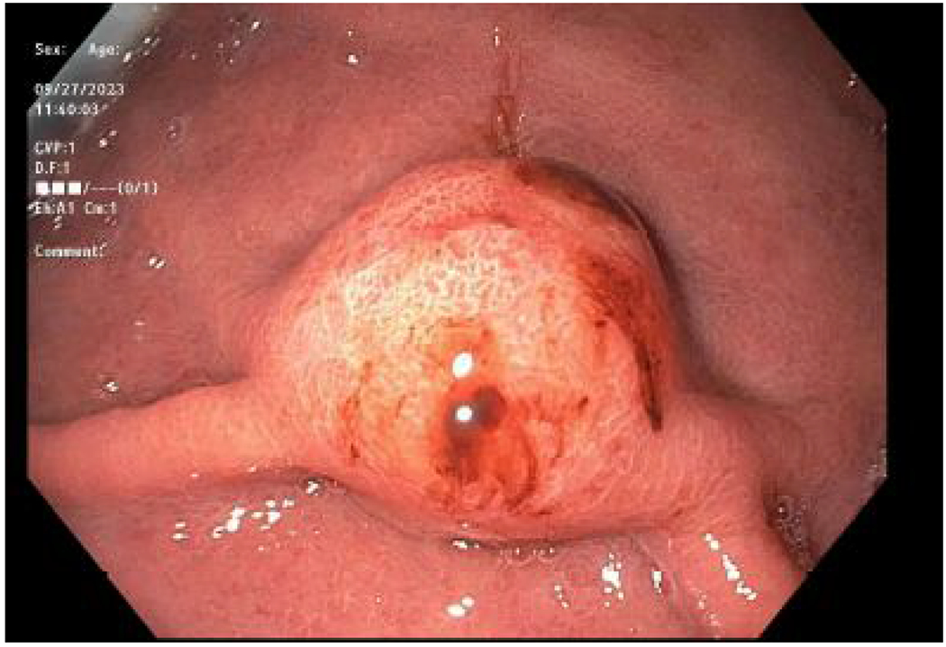
Gastric neuroendocrine tumors (G-NET) are rare tumors arising from enterochromaffin-like cells of the gastric mucosa. They belong to a larger group called gastroenteropancreatic neuroendocrine tumors and are classified as low, intermediate, or high-grade tumors based on their proliferative indices. They are further categorized into three subtypes based on their morphologic characteristics, pathogenesis, and behavior. Types 1 and 2 tumors are characterized by elevated serum gastrin and are usually multifocal. They typically occur in the setting of atrophic gastritis or MEN1/Zollinger Ellison syndrome, respectively. Type 2 tumors are associated with the most symptoms, such as abdominal pain and diarrhea. Type 3 tumors are associated with normal serum gastrin, are usually solitary, and occur sporadically. This type has the most aggressive phenotype and metastatic potential. Treatment and prognosis for G-NET is dependent on their type, size, and stage. Type 1 has the best prognosis, and Type 3 has the worst. This review discusses the presentation, workup, and surgical management of these tumors.
Keywords: GNET; Gastric neuroendocrine; Gastrinoma; High-grade; NET; Neuroendocrine carcinoma.
© 2023. Society of Surgical Oncology.
Figures
Comment in
-
ASO Author Reflections: Simplifying the Fundamentals of Gastric Neuroendocrine Tumor Management.Ann Surg Oncol. 2024 Mar;31(3):1519-1520. doi: 10.1245/s10434-023-14820-6. Epub 2023 Dec 20. Ann Surg Oncol. 2024. PMID: 38123732 No abstract available.
References
-
- Gilligan CJ, Lawton GP, Tang LH, West AB, Modlin IM. Gastric carcinoid tumors: the biology and therapy of an enigmatic and controversial lesion. Am J Gastroenterol. Mar 1995;90(3):338–52. - PubMed
-
- Klimstra DS KG, La Rosa S, Rindi G. Classification of neuroendocrine neoplasms of the digestive system. 5th ed. WHO Classification of Tumours: Digestive System Tumours. International Agency for Research on Cancer; 2019.
Publication types
MeSH terms
Substances
Grants and funding
LinkOut - more resources
Full Text Sources
Medical
