

Preliminary analysis of predicting the first recurrence in patients with neovascular age-related macular degeneration using deep learning
- PMID: 38062449
- PMCID: PMC10702052
- DOI: 10.1186/s12886-023-03229-0
Preliminary analysis of predicting the first recurrence in patients with neovascular age-related macular degeneration using deep learning
Abstract
Background: To predict, using deep learning, the first recurrence in patients with neovascular age-related macular degeneration (nAMD) after three monthly loading injections of intravitreal anti-vascular endothelial growth factor (anti-VEGF).
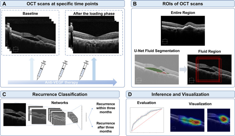
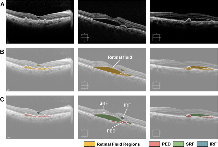
Methods: Optical coherence tomography (OCT) images were obtained at baseline and after the loading phase. The first recurrence was defined as the initial appearance of a new retinal hemorrhage or intra/subretinal fluid accumulation after the initial resolution of exudative changes after three loading injections. Standard U-Net architecture was used to identify the three retinal fluid compartments, which include pigment epithelial detachment, subretinal fluid, and intraretinal fluid. To predict the first recurrence of nAMD, classification learning was conducted to determine whether the first recurrence occurred within three months after the loading phase. The recurrence classification architecture was built using ResNet50. The model with retinal regions of interest of the entire region and fluid region on OCT at baseline and after the loading phase is presented.
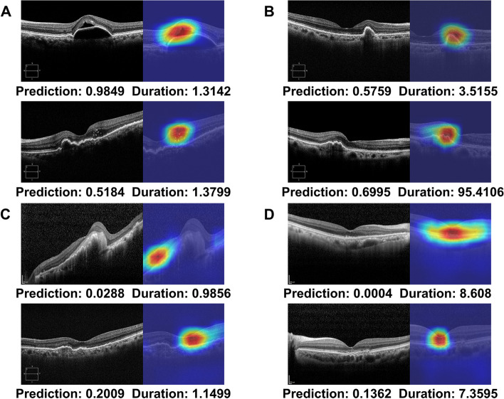
Results: A total of 1,444 eyes of 1,302 patients were included. The mean duration until the first recurrence after the loading phase was 8.20 ± 15.56 months. The recurrence classification system revealed that the model with the fluid region of OCT after the loading phase provided the highest classification performance, with an area under the receiver operating characteristic curve (AUC) of 0.725 ± 0.012. Heatmap analysis revealed that three pathological fluids, subsided choroidal neovascularization lesions, and hyperreflective foci were important areas for the first recurrence.
Conclusions: The deep learning algorithm allowed for the prediction of the first recurrence for three months after the loading phase with adequate feasibility. An automated prediction system may assist in establishing patient-specific treatment plans and the provision of individualized medical care for patients with nAMD.
Keywords: Anti-VEGF; Deep learning; Neovascular age-related macular degeneration; Optical coherence tomography; Recurrence prediction.
© 2023. The Author(s).
Conflict of interest statement
The authors declare no competing interests.
Figures


References
-
- Schmidt-Erfurth U, Eldem B, Guymer R, Korobelnik JF, Schlingemann RO, Axer-Siegel R, Wiedemann P, Simader C, Gekkieva M, Weichselberger A. Efficacy and safety of monthly versus quarterly ranibizumab treatment in neovascular age-related macular degeneration: the EXCITE study. Ophthalmology. 2011;118(5):831–839. doi: 10.1016/j.ophtha.2010.09.004. - DOI - PubMed
MeSH terms
Substances
Grants and funding
- 23ZR1100/Electronics and Telecommunications Research Institute (ETRI) grant funded by the Korean government
- NRF-2021R1F1A1045417/National Research Foundation of Korea (NRF) funded by the Ministry of Science and ICT (Information and Communication Technology)
- 0320222220/Seoul National University Hospital Research Fund
LinkOut - more resources
Full Text Sources
Medical
