. 2023 Dec 8;8(23):e172192.
doi: 10.1172/jci.insight.172192.

Gut mucosal cells transfer α-synuclein to the vagus nerve

Affiliations

Gut mucosal cells transfer α-synuclein to the vagus nerve

Rashmi Chandra et al. JCI Insight. .

Abstract

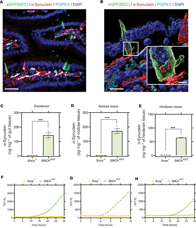
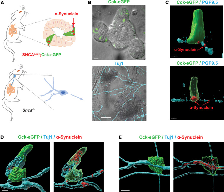
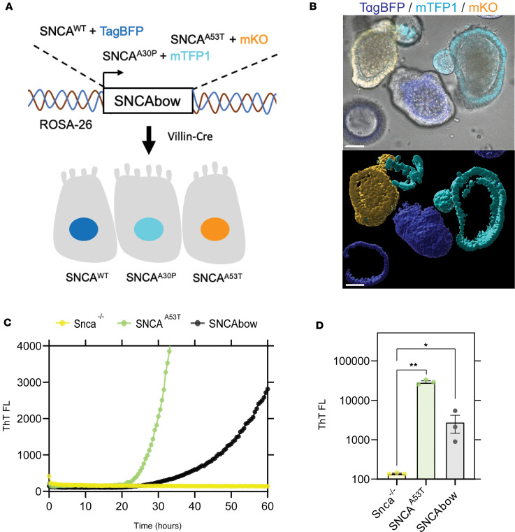
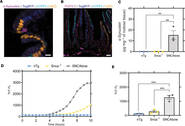
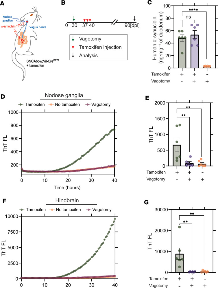
Epidemiological and histopathological findings have raised the possibility that misfolded α-synuclein protein might spread from the gut to the brain and increase the risk of Parkinson's disease. Although past experimental studies in mouse models have relied on gut injections of exogenous recombinant α-synuclein fibrils to study gut-to-brain α-synuclein transfer, the possible origins of misfolded α-synuclein within the gut have remained elusive. We recently demonstrated that sensory cells of intestinal mucosa express α-synuclein. Here, we employed mouse intestinal organoids expressing human α-synuclein to observe the transfer of α-synuclein protein from epithelial cells in organoids to cocultured nodose neurons devoid of α-synuclein. In mice expressing human α-synuclein, but no mouse α-synuclein, α-synuclein fibril-templating activity emerged in α-synuclein-seeded fibril aggregation assays in intestine, vagus nerve, and dorsal motor nucleus. In newly engineered transgenic mice that restrict pathological human α-synuclein expression to intestinal epithelial cells, α-synuclein fibril-templating activity transfered to the vagus nerve and dorsal motor nucleus. Subdiaphragmatic vagotomy prior to induction of α-synuclein expression in intestinal epithelial cells effectively protected the hindbrain from emergence of α-synuclein fibril-templating activity. Overall, these findings highlight a potential non-neuronal source of fibrillar α-synuclein protein that might arise in gut mucosal cells.

Keywords: Gastroenterology; Neuroscience; Parkinson disease.


Figures

Figure 1
Figure 1. α-Synuclein expression and seeding activity in SNCAA53T mice.
(A and B) Immunostaining of duodenum harvested from SNCAA53T mice. Enteroendocrine cells (EECs) expressing green fluorescent protein (shown in green) are scattered among other mucosal cells (DAPI-labeled nuclei, blue) and are in proximity to α-synuclein–containing (red) fibers stained with the pan-neuronal marker PGP9.5 (cyan) in the lamina propria of the villus. ELISA quantification of human α-synuclein in (C) duodenum (α-synuclein quantification in ng/mg of nodose tissue), (D) nodose ganglia (α-synuclein quantification in pg/mg of nodose tissue), and (E) hindbrain (α-synuclein quantification in ng/mg of nodose tissue), from Snca–/– and SNCAA53T mice. RT-QuIC analysis of (F) duodenum, (G) nodose ganglia, and (H) hindbrain of Snca–/– and SNCAA53T mice. Scale bars are 30 μm (A), 10 μm and 1 μm (inset) (B). All the group analyses are shown as mean ± SEM. All RT-QuIC curves shown are representative of the mean from all the groups analyzed. Significance was determined by unpaired t test; ***P < 0.001. n = 3.
Figure 2
Figure 2. Human A53T α-synuclein protein transfers from gut cells to adjoining vagal neurons.
(A) Intestinal organoids were prepared from an SNCAA53T mouse in which CCK-containing cells express enhanced green fluorescent protein (eGFP), and vagal nodose ganglia neurons were isolated from an Snca–/– mouse lacking endogenous α-synuclein. (B) Representative images of organoids and neurons grown in coculture for 5 days, with eGFP-positive cells (green) in the organoid and β-tubulin III (Tuj1, cyan) highlighting neuronal processes. (C) Representative high-magnification α-synuclein (red) staining of an eGFP-positive EEC. Red arrow indicates localization to a PGP9.5-positive (cyan) process in an Snca–/– mouse neuron. (D and E) Representative images with neuron-specific β-tubulin III (cyan). Surface and (adjacent) intracellular confocal slices are shown. Scale bars are 30 μm for B, 3 μm for C, and 5 μm for D and E.
Figure 3
Figure 3. Conditional human α-synuclein expression induces α-synuclein seeding activity in gut organoids.
(A) The SNCAbow expression construct contains 4 tandem cassettes downstream of a chicken β-actin promoter (not shown). The first cassette (not shown) expresses a chemically inducible near-infrared fluorogen-activating peptide (FAP-Mars1). The next 3 cassettes encode a unique fluorescent protein (TagBFP: blue, mTFP1: cyan, or mKO: orange) and a corresponding human synuclein protein SNCAWT, SNCAA30P, and SNCAA53T. When Tg mice are mated to the Villin-Cre (Vil-Cre) strain, Cre-mediated recombination by 3 pairs of orthogonal lox sites (LoxN, Lox2272, LoxP) results in the expression of a single fluorescent protein marker and the corresponding human α-synuclein in any given mucosal cell. (B) Photomicrograph of a small intestine organoid illustrates 3 fluorescent proteins in the mucosa of an SNCAbow mouse indicating the expression of SNCAWT TagBFP (blue), SNCAA30P mTFP1 (turquoise), and SNCAA53T mKO (orange). Scale bar = 30 μm. (C and D) RT-QuIC endpoint ThT fluorescence analysis of nodose ganglia from Snca–/–, SNCAA53T, and SNCAbow mice at 6 months of age. (C) A representative ThT fluorescence profile for these genotypes is provided. (D) Endpoint values were collected after 100 hours of RT-QuIC relative to negative controls. Data were collected and combined from 3 mice for each strain in triplicate. All the group analyses are shown as mean ± SEM. All RT-QuIC curves shown are representative of the mean from all the groups analyzed. Significance was determined by 1-way ANOVA with a Dunnett’s post hoc analysis relative to Snca–/–; *P < 0.05, **P < 0.01, n = 3.
Figure 4
Figure 4. Conditional human α-synuclein expression in gut mucosal cells produces in α-synuclein seeding activity in nodose ganglia.
(A) SNCAbow mouse duodenum illustrating expression of fluorescent protein markers BFP (blue), TFP (turquoise), and mKO2 (orange) in mucosal cells (enterocytes and EECs). α-Synuclein (magenta) immunostaining is present in a single intestinal mucosal cell consistent with an EEC (scale bar = 10 μm). (B) Duodenum from SNCAbow mouse illustrating PGP9.5-positive neuronal fibers (magenta) innervating intestinal crypts and villi (scale bar = 50 μm). (C) ELISA quantification of human α-synuclein protein in nodose ganglia of nontransgenic, Snca–/–, and SNCAbow mice. (D) A representative ThT fluorescence profile (RT-QuIC) and endpoint analysis of nodose ganglia from nontransgenic (nTg), Snca–/–, and SNCAbow mice at 1 month of age. (E) RT-QuIC analysis of nodose ganglia from 6-month-old nTg, Snca–/–, and SNCAbow mice. All the group analyses are shown as mean ± SEM. All RT-QuIC curves shown are representative of the mean from all the groups analyzed. Significance was determined by 1-way ANOVA with a Dunnett’s post hoc analysis relative to SNCAbow; **P < 0.01, ***P < 0.001, n = 3.
Figure 5
Figure 5. Vagotomy spares the nodose ganglia from α-synuclein seeding activity and prevents spread to the hindbrain.
(A and B) Experimental model. SNCAbow Vil-CreERT2 mice underwent bilateral subdiaphragmatic vagotomy or sham surgery 1 week before tamoxifen treatment. (C) ELISA measurements of α-synuclein protein in the gut 3 months after tamoxifen treatment. RT-QuIC analysis of (D and E) vagal nodose ganglia and (F and G) hindbrain analyzed 3 months after tamoxifen treatment. Representative ThT fluorescence profiles are shown in D and F. All the group analyses are shown as mean ± SEM. All RT-QuIC curves shown are representative of the mean from all the groups analyzed. Data points in B, E, and G represent the significance determined by a 1-way ANOVA with a Dunnett’s post hoc analysis relative to tamoxifen-treated and no-vagotomy groups. **P < 0.01, ****P < 0.0001, n = 6.

Update of

References

    1. Chaudhuri KR, Schapira AH. Non-motor symptoms of Parkinson’s disease: dopaminergic pathophysiology and treatment. Lancet Neurol. 2009;8(5):464–474. doi: 10.1016/S1474-4422(09)70068-7. - DOI - PubMed
    1. Bisaglia M, et al. Structural insights on physiological functions and pathological effects of alpha-synuclein. FASEB J. 2009;23(2):329–340. doi: 10.1096/fj.08-119784. - DOI - PubMed
    1. Soto C. Transmissible proteins: expanding the prion heresy. Cell. 2012;149(5):968–977. doi: 10.1016/j.cell.2012.05.007. - DOI - PMC - PubMed
    1. Bendor JT, et al. The function of α-synuclein. Neuron. 2013;79(6):1044–1066. doi: 10.1016/j.neuron.2013.09.004. - DOI - PMC - PubMed
    1. Polymeropoulos MH, et al. Mutation in the alpha-synuclein gene identified in families with Parkinson’s disease. Science. 1997;276(5321):2045–2047. doi: 10.1126/science.276.5321.2045. - DOI - PubMed

Publication types

Substances