. 2023 Dec 8;14(1):8143.
doi: 10.1038/s41467-023-43887-8.

Structural plasticity for neuromorphic networks with electropolymerized dendritic PEDOT connections

Affiliations

Structural plasticity for neuromorphic networks with electropolymerized dendritic PEDOT connections

Kamila Janzakova et al. Nat Commun. .

Abstract

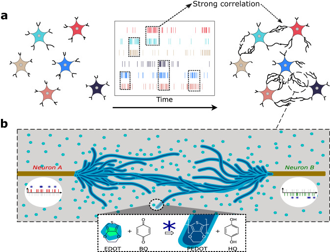
Neural networks are powerful tools for solving complex problems, but finding the right network topology for a given task remains an open question. Biology uses neurogenesis and structural plasticity to solve this problem. Advanced neural network algorithms are mostly relying on synaptic plasticity and learning. The main limitation in reconciling these two approaches is the lack of a viable hardware solution that could reproduce the bottom-up development of biological neural networks. Here, we show how the dendritic growth of PEDOT:PSS-based fibers through AC electropolymerization can implement structural plasticity during network development. We find that this strategy follows Hebbian principles and is able to define topologies that leverage better computing performances with sparse synaptic connectivity for solving non-trivial tasks. This approach is validated in software simulation, and offers up to 61% better network sparsity on classification and 50% in signal reconstruction tasks.


Conflict of interest statement

The authors declare no competing interests.

Figures

Fig. 1
Fig. 1. Structural plasticity emulation with PEDOT:PSS dendritic fibers.
a As the activity correlates between every pair of neurons, the dendrites between them grows, creating a physical topology of artificial neural networks. b Dendritic electropolymerization is obtained by applying AC electrical signal in between conducting Au wires. Electrical potential at each node drives oxidation of EDOT molecules into PEDOT and reduction of benzoquinone (BQ) into hydroquinone (HQ). A pulse of voltage on each terminal represents node activity that can correlate to create overpotential (blue star), leading to electropolymerization.
Fig. 2
Fig. 2. Dendrites electropolymerization induced by spike timing modulation.
a Setup for dendrites electropolymerization induced by spike-timing activity of applied signals. Bipolar pulses with opposite polarity were applied from both electrodes to emulate activity of neurons with time difference in between both of ΔT. b Optical microscope images of completed dendritic branches obtained when ΔT time shift increases. Bipolar pulses are repeated at frequency fpre=fpost {20 Hz, 80 Hz and 130 Hz}. No connections occurred at ΔT = 1.5 ms and 2 ms for F = 20 Hz. c Longitudinal growth rate evaluation and d conductance evolution of dendritic connections synthesized at different ΔT values and at fpre=fpost {20 Hz, 80 Hz and 130 Hz}. Growth rate is evaluated by considering a gap distance of 240 μm and a completion time corresponding to the time when dendrites of both electrodes first connect to each other. Note that after bridging branches with each other, the dendritic growth saturates, and no further significant changes in the number of branches and branches diameter were observed (see supplementary, Figs. S11 and S12). Error bars in c are represented by the standard deviation expressed in percentage.
Fig. 3
Fig. 3. Pavlovian learning promoted by means of spike-timing dependent activity electropolymerization conducted in two phases.
a Neuron-level schematic representing the growable connectivity between each pair of neurons, associated with food, saliva and bell. b Time-lapse of dendritic growth during 1st phase in which time correlation and corresponding overpotentiation took place only between “food" and “saliva" pulses (signifying that salivation naturally occurs only as response to the food stimuli). c Dendritic evolution with time during 2nd phase in which as a result of associative learning the correlated activity is as well introduced for “bell" and “saliva" signals leading to pairing of these nodes by polymer fibers.
Fig. 4
Fig. 4. Dendritic growth from data to model.
a Time-lapse of physical dendritic growth on four electrodes with unlike frequencies. Longitudinal growth rate (b) and conductance (c) of dendritic connections between wires stimulated by random Poisson-distributed pulses of various frequencies Fi and Fj, compared with the dendritic growth model.
Fig. 5
Fig. 5. Electromyography classification task.
a Schematic overview of the EMG classification task. Each input of the eight channels is first spike-encoded into 16 spike trains. The resulting spike activity is passed through the structural plasticity model with three output neurons, creating the 16→3 connectivity matrix. The output neurons spikes at either 1000 or 100 Hz, associated with the correct and incorrect classes, respectively, of the currently presented label. b Resulting topologies of the dendritic growth stopped at various points in time. Partial connections are indicated in green, and fully connected nodes are linked by a gold-colored link. The organization of the nodes is schematic and does not reflect the actual placement of electrodes. c Two different networks were tested with modeled structural plasticity data. In the first model, the spikes go through the sparse connectivity matrix with binary weights, and spike counts are accumulated before being classified with a SVM. In the second model, spikes are accumulated directly on the 16 inputs, and a sparse weighted tensor is trained with stochastic gradient descent. Weight training is done after the creation of the connectivity matrix using structural plasticity in all cases. d Test accuracy of the EMG task as a function of the total number of connections. The two training methods are compared with a random topology with random initial weights, vs the structural plasticity-based topology. Ten different random topologies are generated and averaged for the random connectivity. Standard deviation is presented with a shaded area for each corresponding curve.
Fig. 6
Fig. 6. Sparse unsupervised spike auto-encoding task.
a Schematic overview of the spike auto-encoding task. A random multi-channels input signal is connected by a linear input layer and fed to a pool of recurrent spiking neurons as currents. The spikes of the reservoir are filtered and decoded by an output layer to retrieve the original signal. The input layer is fixed and sparsely connected to the recurrent layer called the reservoir. The reservoir layer connectivity matrix is generated by either the structural plasticity method, or with random connectivity. b Two-dimensional lattice graph of spiking neurons at different point in time during the growth of connections with the structural plasticity model. The nodes' organization is simulated and does not depict the position of electrodes in a physical system. Each pair of neuron can grow a connection in-between. Green links represent partial connections, and gold-colored links are fully created connections. c Sparse unsupervised spike encoding error as a function of the connection density. The connection density represents the number of connections divided by the total possible number of connections. Self-connections are not included. The resulting mean squared error for encoding a random white-noise signal is reported. In the left plot, random connectivity matrices are generated for various target densities. In the right plot, the structural plasticity software model was used with different parameters (pulse duration and interneuron distance) with a fixed two-second duration as to create topologies with various densities.

References

    1. Indiveri G, et al. Neuromorphic silicon neuron circuits. Front. Neurosci. 2011;5:73. doi: 10.3389/fnins.2011.00073. - DOI - PMC - PubMed
    1. Sangwan VK, Hersam MC. Neuromorphic nanoelectronic materials. Nat. Nanotechnol. 2020;15:517–528. doi: 10.1038/s41565-020-0647-z. - DOI - PubMed
    1. Tang J, et al. Bridging Biological and Artificial Neural Networks with Emerging Neuromorphic Devices: Fundamentals, Progress, and Challenges. Adv. Mater. 2019;31:e1902761. doi: 10.1002/adma.201902761. - DOI - PubMed
    1. Frémaux N, Gerstner W. Neuromodulated Spike-Timing-Dependent Plasticity, and Theory of Three-Factor Learning Rules. Front. Neural Circuits. 2015;9:85. - PMC - PubMed
    1. Martin E, et al. EqSpike: spike-driven equilibrium propagation for neuromorphic implementations. iScience. 2021;24:102222. doi: 10.1016/j.isci.2021.102222. - DOI - PMC - PubMed