. 2023 Dec 7;13(1):21698.
doi: 10.1038/s41598-023-48572-w.

Correlation of PD-1 and PD-L1 expression in oral leukoplakia and oral squamous cell carcinoma: an immunohistochemical study

Affiliations

Correlation of PD-1 and PD-L1 expression in oral leukoplakia and oral squamous cell carcinoma: an immunohistochemical study

L R Greeshma et al. Sci Rep. .

Abstract

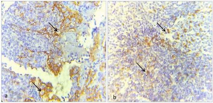
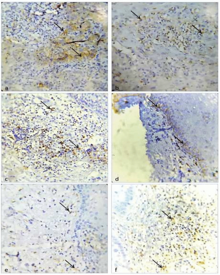
The programmed cell death protein (PD-1)/programmed cell death protein ligand (PD-L1) pathway and cytotoxic T lymphocyte antigen are the most important co-stimulatory molecules that play a key role in the negative regulation of T cells during carcinogenesis. We aimed to evaluate the immunohistochemical expression of PD-1 and PD-L1 in oral leukoplakia and squamous cell carcinoma compared with normal oral mucosa. Twenty-five cases of oral squamous cell carcinoma, oral leukoplakia and normal oral mucosa tissue specimens were immunohistochemically stained to assess PD-1 and PD-L1 expression. The PD-L1 positivity of subepithelial TAFs (p < 0.001) increased with increasing grades of oral leukoplakia. Pearson's correlation indicated a high positive correlation between the PD-L1 labelling index of epithelial tumour cells and the PD-1 labelling index of tumour infiltrating lymphocytes (p value: 0.005) in OSCC. A high positive correlation was noted between the H-score of PD-L1 positive tumour epithelial cells and the H-score of PD-1 positive tumour infiltrating lymphocytes in OSCC (p value: 0.001). PD-L1 positivity increased in dysplastic epithelial cells from premalignant lesions to malignancy. The sub-epithelial PD-L1 positive TAFs were higher in oral leukoplakia compared to OSCC inferring that PD-L1 positivity in TAFs decreased with malignant transformation. The PD-1 positivity in TILs was higher in oral leukoplakia than in OSCC.


Conflict of interest statement

The authors declare no competing interests.

Figures

Figure 1
Figure 1
Photo micrograph showing immunohistochemical expression. (a) Tonsillar epithelium as positive control of PD-L1 (400×) (black arrow heads). (b) Tonsillar lymphocytesas positive control for PD-1 (400×) (black arrow heads).
Figure 2
Figure 2
Photo micrograph showing immunohistochemical expression. (a) Membranous expression of PD-L1 in epithelial tumour cells present in the invasive front of oral squamous cell carcinoma (400×) (black arrow heads). (b) Membranous expression of PD-L1 in tumour associated fibroblast cells present in the invasive front of oral squamous cell carcinoma (400×) (black arrow heads). (c) Membranous expression of PD-1 in tumour- infiltrating lymphocytes (TILs) present in the invasive front of oral squamous cell carcinoma (400×) (black arrow heads). (d) Membranous expression of PD-L1 in dysplastic basal epithelial cells of oral leukoplakia (400×) (black arrow heads). (e) Membranous expression of PD-L1 in tumour associated fibroblast cells present subepithelial stroma of oral leukoplakia (400×) (black arrow heads). (f) Membranous expression of PD-1 infiltrating lymphocytes present in subepithelial stroma of oral leukoplakia (400×) (black arrow heads).
Figure 3
Figure 3
(a) Photo micrograph showing immunonegativity for PD-L1 in Normal Oral Mucosa. (b) Photo micrograph showing immunonegativity for PD-1 in Normal Oral Mucosa.

References

    1. Warnakulasuriya S. Living with oral cancer: Epidemiology with particular reference to prevalence and life-style changes that influence survival. Oral Oncol. 2010;46(6):407–410. doi: 10.1016/j.oraloncology.2010.02.015. - DOI - PubMed
    1. Rhodus NL, Kerr AR, Patel K. Oral cancer: Leukoplakia, premalignancy, and squamous cell carcinoma. Dent. Clin. N. Am. 2014;58(2):315–340. doi: 10.1016/j.cden.2013.12.004. - DOI - PubMed
    1. Moro JD, Maroneze MC, Ardenghi TM, Barin LM, Danesi CC. Oral and oropharyngeal cancer: Epidemiology and survival analysis. Einstein (São Paulo) 2018;16(2):eAO4248. doi: 10.1590/s1679-45082018ao4248. - DOI - PMC - PubMed
    1. Feller L, Lemmer J. Oral squamous cell carcinoma: Epidemiology, clinical presentation and treatment. J. Cancer Ther. 2012;1:03.
    1. Warnakulasuriya S, Johnson NW, Van der Waal I. Nomenclature and classification of potentially malignant disorders of the oral mucosa. J. Oral Pathol. Med. 2007;36(10):575–580. doi: 10.1111/j.1600-0714.2007.00582.x. - DOI - PubMed

MeSH terms

Substances