. 2024 Apr;45(4):815-830.
doi: 10.1038/s41401-023-01193-5. Epub 2023 Dec 8.

Lonicerin promotes wound healing in diabetic rats by enhancing blood vessel regeneration through Sirt1-mediated autophagy

Affiliations

Lonicerin promotes wound healing in diabetic rats by enhancing blood vessel regeneration through Sirt1-mediated autophagy

Zhen Lin et al. Acta Pharmacol Sin. 2024 Apr.

Abstract

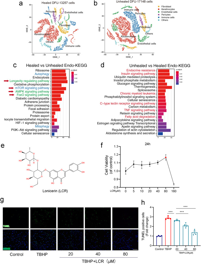
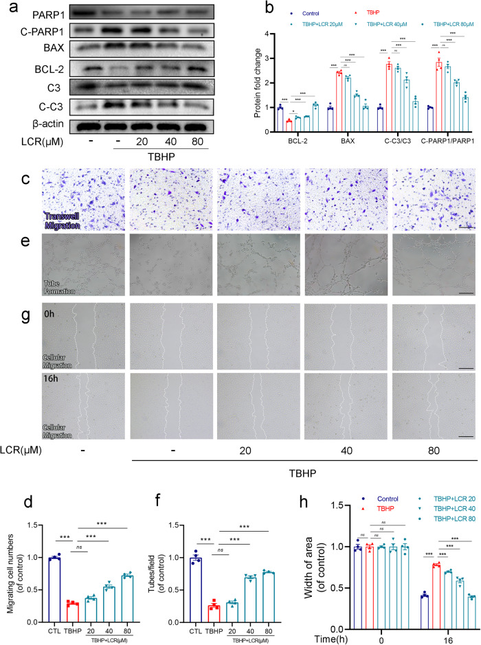
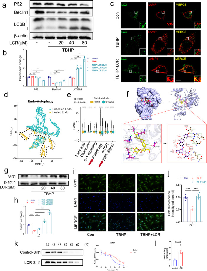
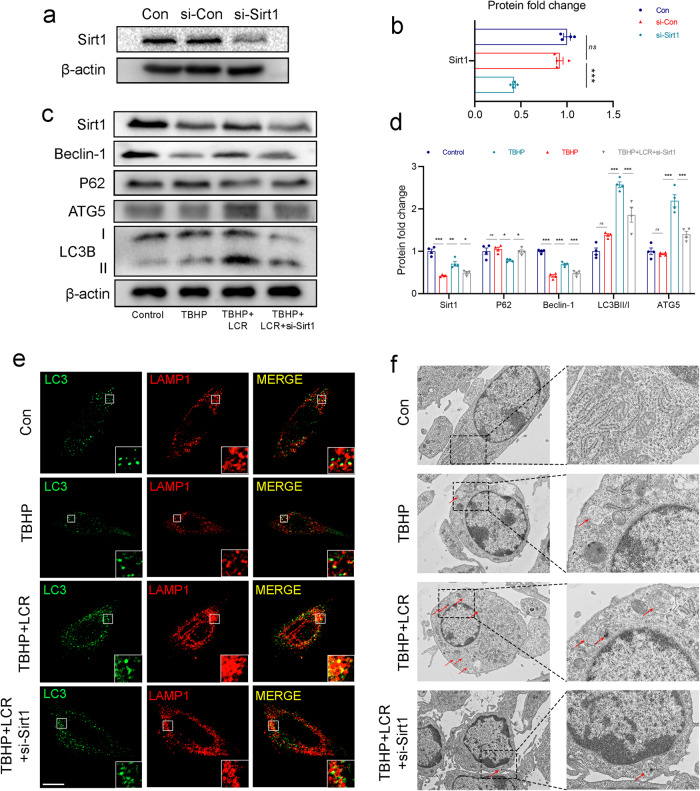
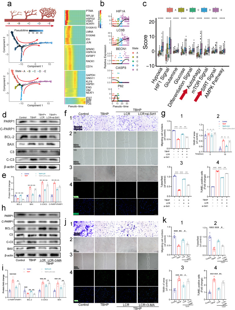
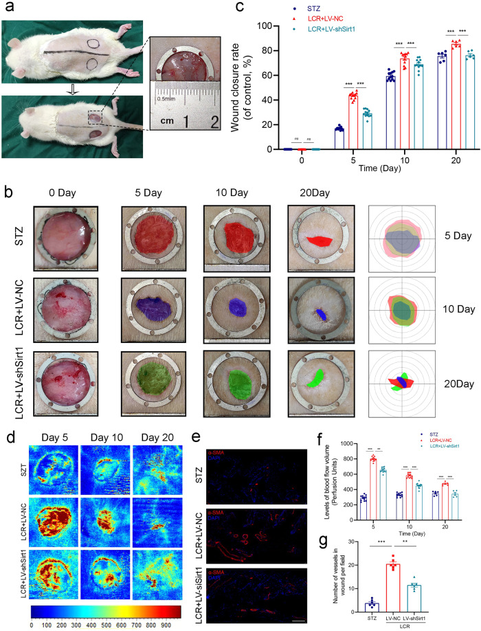
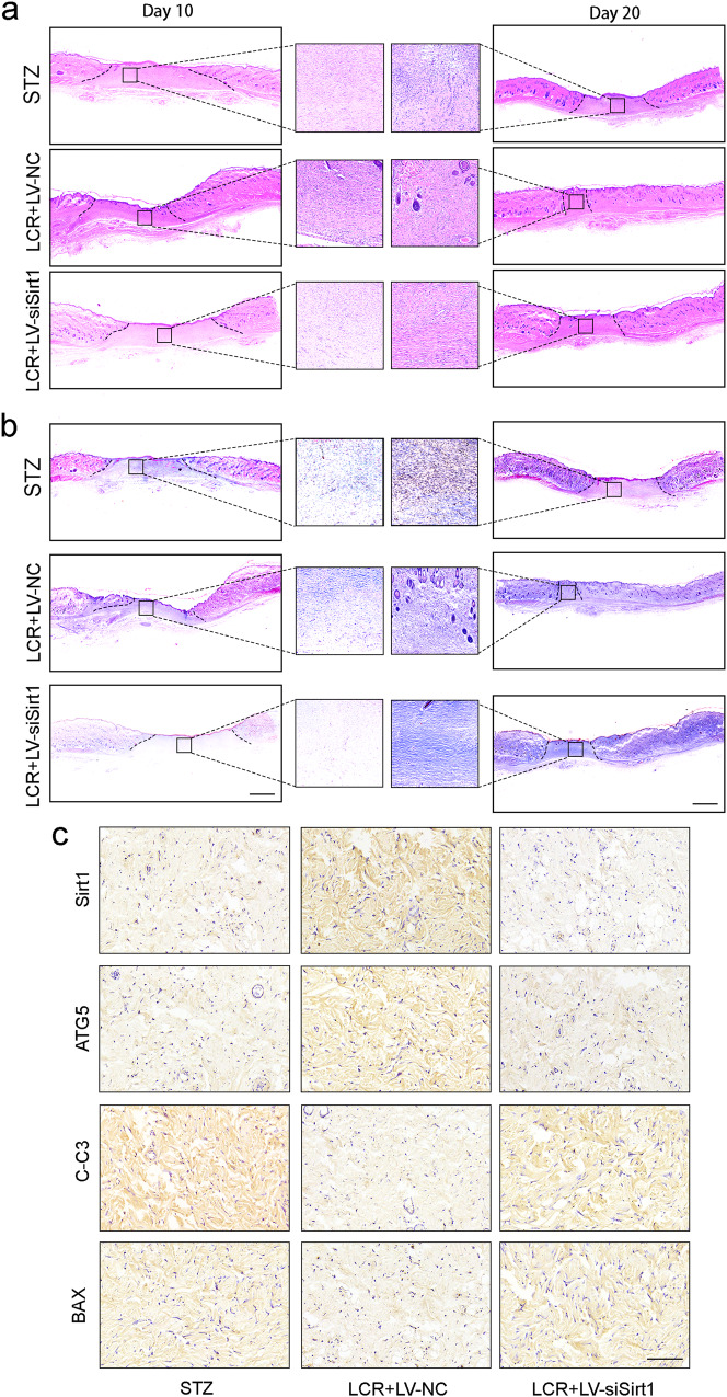
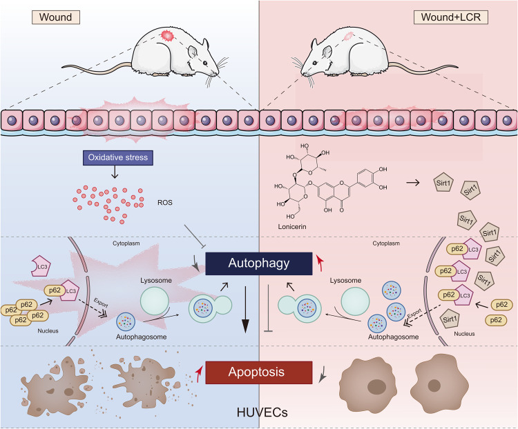
Among the numerous complications of diabetes mellitus, diabetic wounds seriously affect patients' quality of life and result in considerable psychological distress. Promoting blood vessel regeneration in wounds is a crucial step in wound healing. Lonicerin (LCR), a bioactive compound found in plants of the Lonicera japonica species and other honeysuckle plants, exhibits anti-inflammatory and antioxidant activities, and it recently has been found to alleviate ulcerative colitis by enhancing autophagy. In this study we investigated the efficacy of LCR in treatment of diabetic wounds and the underlying mechanisms. By comparing the single-cell transcriptomic data from healing and non-healing states in diabetic foot ulcers (DFU) of 5 patients, we found that autophagy and SIRT signaling activation played a crucial role in mitigating inflammation and oxidative stress, and promoting cell survival in wound healing processes. In TBHP-treated human umbilical vein endothelial cells (HUVECs), we showed that LCR alleviated cell apoptosis, and enhanced the cell viability, migration and angiogenesis. Furthermore, we demonstrated that LCR treatment dose-dependently promoted autophagy in TBHP-treated HUVECs by upregulating Sirt1 expression, and exerted its anti-apoptotic effect through the Sirt1-autophagy axis. Knockdown of Sirt1 significantly decreased the level of autophagy, and mitigated the anti-apoptotic effect of LCR. In a STZ-induced diabetic rat model, administration of LCR significantly promoted wound healing, which was significantly attenuated by Sirt1 knockdown. This study highlights the potential of LCR as a therapeutic agent for the treatment of diabetic wounds and provides insights into the molecular mechanisms underlying its effects.

Keywords: Sirt1; angiogenesis; autophagy; diabetic wounds; lonicerin; oxidative stress; wound healing.


Conflict of interest statement

The authors declare no competing interests.

Figures

Fig. 1
Fig. 1. Lonicerin treatment decreases TBHP-induced apoptosis in HUVECs.
a, b tSNE plot showing clusters and annotations of cells identified in patients with Healed DFU and Unhealed DFU, respectively. Cells from different patients gather together. Different cell types are shown in different colors; (c, d) KEGG enrichment analysis of the genes in endothelial cells in patients with Healed DFU and Unhealed DFU, respectively; (e) The chemical structure for LCR; (f) CCK8 assay of HUVECs was conducted to measure relative cell viability after being treated with different concentrations of LCR; (g, h) Representative images demonstrating terminal deoxynucleotidyl transferase deoxyuridine triphosphate nick end labeling-positive nuclei (green color). Scale bar = 100 μm. HUVECs treated with or without LCR for 24 h. The values presented are the means ± SEM (n = 4). < 0.05, P < 0.01, ***P < 0.001.
Fig. 2
Fig. 2. Lonicerin combats TBHP-induced apoptosis and promotes the function of HUVECs.
a, b The protein expression of BCL-2, BAX, C-PARP1/PARP and C-C3/C3 was detected by Western blot in the HUVECs; (c, d) Transwell migration assay results demonstrate the effect of LCR on HUVEC migration. Scale bar = 100 μm; (e, f) Tube formation assay results demonstrate the effect of LCR on HUVEC neovascularization. Scale bar = 100 μm; (g, h) Representative images of scratch wound healing assay of HUVECs in vitro at 0 and 16 h posttreatment and its quantitative analysis. Scale bar = 100 μm. HUVECs treated with or without 20, 40, 80 μM LCR for 24 h. The values presented are the means ± SEM (n = 4). **P < 0.01, ***P < 0.001. ns indicates no significant difference.
Fig. 3
Fig. 3. Lonicerin promotes autophagy in TBHP-treated HUVECs.
a, b The protein expression of P62, Beclin-1, LC3B in HUVECs treated with different concentrations of LCR was visualized by Western blotting; (c) The double immunofluorescence staining of LC3B and LAMP1 in TBHP-exposed HUVECs treated with LCR (green signals represent LC3B), red signals represent LAMP1, scale bar = 10 μm; (d) tSNE plot showing clusters of endothelial cells in autophagy. Different patient groups are shown in different colors. Blue represents the endothelial cells of unhealed patients, yellow represents the endothelial cells of healed patients; (e) Scores for autophagy, SIRT signal, fatty acid and glutamine metabolism, and IL-1 signaling calculated by PCA method in keratinocytes comparing healed patients versus unhealed patients; (f) The 3D and 2D results of the optimal conformation of LCR binding to Sirt1 showed that the linked amino acids were ALA-204, LYS-203, ARG-274, ALA-262, GLN-345 and VAL-412, respectively; (g, h) The protein expression of Sirt1 was detected by Western blot in the HUVECs; (i) Immunofluorescence staining of Sirt1 (green). Nuclei were counterstained with DAPI (blue) scale bar = 20 μm; (j) Quantification data of mean fluorescence intensity of Sirt1; (k) In the CETSA assay, the degree of protein expression of Sirt1 in HUVECs in the presence and absence of LCR; (l) The effect of LCR on Sirt1 activity was detected by Sirt1 activity assay kit. HUVECs treated with or without 20, 40, 80 μM LCR for 24 h. The values presented are the means ± SEM (n = 4). **P < 0.01, ***P < 0.001.
Fig. 4
Fig. 4. Lonicerin promotes autophagy by increasing Sirt1 expression.
a, b The knockdown effect of si-RNA was tested by Western blot; (c, d) The Western blot results of Sirt1, Beclin-1, P62, ATG5 and LC3B under LCR treatment with si-Sirt1 before TBHP addition; (e) The representative images of double immunofluorescence staining in HUVECs treated as above (green signals represent LC3B), red signals represent LAMP1, scale bar = 10 μm; (f) TEM images of autophagic vesicles in HUVECs treated as indicated (red arrow: autophagic vesicles). Scale bar = 1 μm. HUVECs treated with or without 80 μM LCR for 24 h. The values presented are the means ± SEM (n = 4). *P < 0.05, **P < 0.01, ***P < 0.001. ns indicates no significant difference.
Fig. 5
Fig. 5. Anti-apoptotic effects of Lonicerin (LCR) are related to Sirt1 in HUVECs.
a Pseudotime analysis showed the endothelial cell development pattern; (b) HIF1A, LC3B, and BECN1 increased, whereas CASP3 of endothelial cells decreased, but p62 increased firstly and then down with pseudotime; (c) The gene expression scores for hypoxia, glutamine, and glucose metabolism, and autophagy, AMPK, SIRT, HIF1, differentiation signaling pathways as divided by wound healing stage. Box plot graphs indicated the value of minimum, first, quartile, median, third quartile, and maximum; (d, e) The Western blot results of BCL-2, BAX, C-PARP1/PARP1 and C-C3/C3 under LCR treatment with si-Sirt1 pretreatment before TBHP addition; (f, g, 1) Representative transwell migration assay images of HUVECS treated with LCR and si-Sirt1 pretreatment before TBHP addition. Scale bar = 100 μm; (f, g, 2) Representative images of scratch wound healing assay of HUVECs in vitro at 0 and 16 h posttreatment and its quantitative analysis. Scale bar = 100 μm; (f, g, 3) Tube formation assay results demonstrate the effect of LCR on HUVEC neovascularization. Scale bar, 100 μm; (f, g, 4) Apoptotic chondrocytes were examined using TUNEL fluorescence immunocytochemistry (green). Nuclei were counterstained with DAPI (blue). Scale bar = 100 μm; (h, i) The Western blot results of BCL-2, BAX, C-PARP1/PARP1 and C-C3/C3 under LCR treatment with 3-MA pretreatment before TBHP addition; (j, k, 1) Representative transwell migration assay images of HUVECs treated with LCR and 3-MA pretreatment before TBHP addition. Scale bar = 100 μm; (j, k, 2) Tube formation assay results demonstrate the effect of LCR on HUVEC neovascularization. Scale bar, 100 μm; (j, k, 3) Representative images of scratch wound healing assay of HUVECs in vitro at 16 h posttreatment and its quantitative analysis. Scale bar = 100 μm; (j, k, 4) Apoptotic chondrocytes were examined using TUNEL fluorescence immunocytochemistry (green). Nuclei were counterstained with DAPI (blue). Scale bar = 100 μm; HUVECs treated with or without 80 μM LCR for 24 h. The values presented are the means ± SEM (n = 4). *P < 0.05, **P < 0.01, ***P < 0.001. ns indicates no significant difference.
Fig. 6
Fig. 6. Lonicerin accelerates cutaneous wound healing in rats.
a As the animal experimental schematics show, rats were anesthetized and the dorsal fur was shaved and then two full-thickness wounds were made with biopsy punch per mouse; (b) Representative images of the mice skin wound healing in the STZ group, LCR + LV-NC group, and LCR + LV-shSirt1 groups. The rulers in the pictures are shown in millimeters; (c) Wound area closure rate at different time points (days 0, 5, 10 and 20) in groups mentioned above. n = 12 or 6 independent animals per group; (d, f) Representative color laser Doppler images are taken to value subcutaneous blood flow on postsurgery days 5, 10 and 20. n = 12 or 6 independent animals per group. n = 12 or 6 independent animals per group; (e, g) Immunofluorescence staining for α-SMA (red) and their quantified results (n = 6). Scale bar = 100 μm; The values presented are the means ± SEM. **P < 0.01, ***P < 0.001. ns indicates no significant difference. ns indicates no significant difference.
Fig. 7
Fig. 7. Lonicerin accelerates cutaneous wound healing in rats (continue).
a Representative H&E-stained sections of dorsal skin wound samples from different groups on day 10 and day 20. Black solid line indicated an epidermal gap. Scale bar = 500 μm; (b) Representative Masson’s trichrome-stained sections of dorsal skin wound samples from different groups on day 10 and day 20. Scale bar = 500 μm; (c) Immunohistochemistry staining images of Sirt1, ATG5, C-C3 and BAX of the control, LCR + LV-NC, and LCR + LV-siSirt1 groups. Scale bar = 100 μm. n = 6 independent animals per group.
Fig. 8
Fig. 8
Lonicerin promotes diabetic wound healing by enhancing blood vessel regeneration through Sirt1-mediated autophagy.

References

    1. The Lancet Diabetes E. Diabetes: mapping the titanic struggle ahead. The Lancet Diabetes Endocrinol. 2018;6:1. doi: 10.1016/S2213-8587(17)30414-X. - DOI - PubMed
    1. Long Y, Wei H, Li J, Yao G, Yu B, Ni D, et al. Effective wound healing enabled by discrete alternative electric fields from wearable nanogenerators. ACS Nano. 2018;12:12533–40. doi: 10.1021/acsnano.8b07038. - DOI - PMC - PubMed
    1. Shiekh PA, Singh A, Kumar A. Exosome laden oxygen releasing antioxidant and antibacterial cryogel wound dressing oxoband alleviate diabetic and infectious wound healing. Biomaterials. 2020;249:120020. doi: 10.1016/j.biomaterials.2020.120020. - DOI - PubMed
    1. Martí-Carvajal AJ, Gluud C, Nicola S, Simancas-Racines D, Reveiz L, Oliva P, et al. Growth factors for treating diabetic foot ulcers. Cochrane Database Syst Rev. 2015;2015:Cd008548. - PMC - PubMed
    1. Chang M, Nguyen TT. Strategy for treatment of infected diabetic foot ulcers. Acc Chem Res. 2021;54:1080–93. doi: 10.1021/acs.accounts.0c00864. - DOI - PubMed

MeSH terms

LinkOut - more resources