. 2023 Nov 25;13(23):3530.
doi: 10.3390/diagnostics13233530.

Diagnostic Challenges in ABCA4-Associated Retinal Degeneration: One Gene, Many Phenotypes

Affiliations

Diagnostic Challenges in ABCA4-Associated Retinal Degeneration: One Gene, Many Phenotypes

Tien-En Tan et al. Diagnostics (Basel). .

Abstract

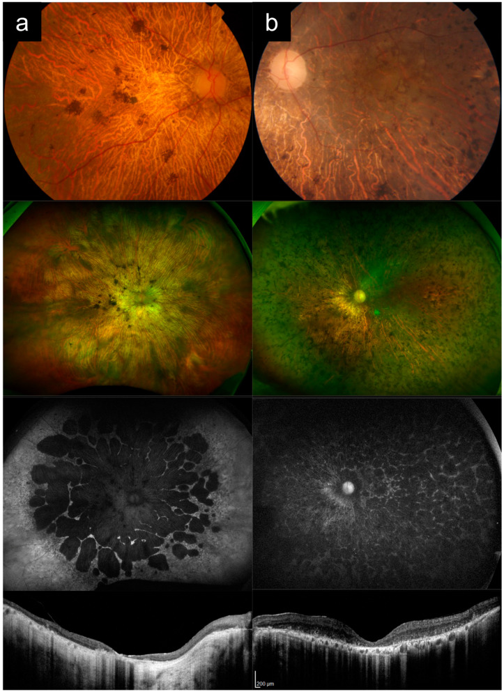
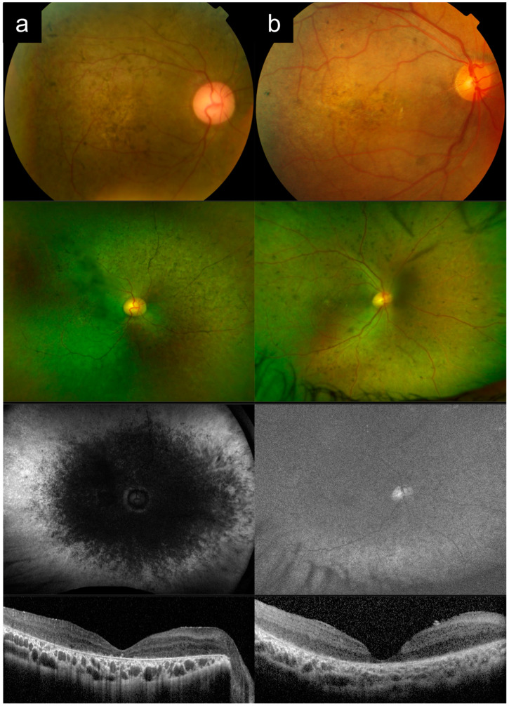
(1) Purpose: ABCA4-associated retinal degeneration (ABCA4-RD) is a phenotypically diverse disease that often evades diagnosis, even by experienced retinal specialists. This may lead to inappropriate management, delayed genetic testing, or inaccurate interpretation of genetic testing results. Here, we illustrate the phenotypic diversity of ABCA4-RD using a series of representative cases and compare these to other conditions that closely mimic ABCA4-RD. (2) Methods: Genetically confirmed ABCA4-RD cases with representative phenotypes were selected from an inherited retinal disease cohort in Singapore and compared to phenocopies involving other retinal diseases. (3) Results: ABCA4-RD phenotypes in this series included typical adolescent-onset Stargardt disease with flecks, bull's eye maculopathy without flecks, fundus flavimaculatus, late-onset Stargardt disease, and severe early-onset Stargardt disease. Phenocopies of ABCA4-RD in this series included macular dystrophy, pattern dystrophy, cone dystrophy, advanced retinitis pigmentosa, Leber congenital amaurosis, drug toxicity, and age-related macular degeneration. Key distinguishing features that often suggested a diagnosis of ABCA4-RD were the presence of peripapillary sparing, macular involvement and centrifugal distribution, and a recessive pedigree. (4) Conclusions: ABCA4-RD demonstrates a remarkable phenotypic spectrum that makes diagnosis challenging. Awareness of the clinical spectrum of disease can facilitate prompt recognition and accurate diagnostic testing.

Keywords: ABCA4; Stargardt disease; genetic diagnosis; inherited retinal disease; phenotypic variation; retinal dystrophy.


Conflict of interest statement

The authors declare no conflict of interest.

Figures

Figure 1
Figure 1
Cases 1a versus 1b: Typical adolescent-onset Stargardt disease with macular atrophy, flecks and peripapillary sparing (a) versus PRPH2-associated macular dystrophy (b). Note the similar appearance on autofluorescence imaging, except for the peripapillary sparing, which is present in Case 1a, but absent in Case 1b. These cases can also be distinguished clinically based on age of onset and inheritance patterns.
Figure 2
Figure 2
Cases 2a versus 2b versus 2c: Stargardt disease without flecks (a) versus GUCY2D-associated cone dystrophy (b) versus hydroxychloroquine toxicity (c). Note the similar appearance between Cases 2a and Case 2b, particularly on autofluorescence and optical coherence tomography imaging. The slight metallic sheen within the area of atrophy on the color photograph in Case 2a points towards ABCA4-associated Stargardt disease. These cases can also be distinguished based on clinical symptoms, electroretinography, and inheritance patterns. Case 2c shows that systemic drug toxicity can present with similar imaging features that mimic inherited retinal disease.
Figure 3
Figure 3
Cases 3a versus 3a: Fundus flavimaculatus (a) versus autosomal-dominant pattern dystrophy (b). These cases both presented with late onset, yellow flecks, good vision, and minimal macular atrophy. Note the peripapillary sparing on autofluorescence imaging in Case 3a that points towards ABCA4-associated retinal degeneration. Also note the prominent pigment clumps within the yellow flecks in Case 3b, which are more characteristic of PRPH2-associated pattern dystrophy, although genetic testing results were not available for this case. These cases can also be distinguished based on the inheritance patterns.
Figure 4
Figure 4
Cases 4a versus 4b: Late-onset Stargardt disease (a) versus dry age-related macular degeneration (AMD) with geographic atrophy (b). In Case 4a, the yellow flecks are more elongated and pisciform, and many more flecks are apparent on autofluorescence imaging where they are both hyper- and hypo-autofluorescent. There is also peripapillary sparing, with the nasal edge of the atrophy concave towards the disc as a result. In Case 4b, the yellow drusen are rounder, and do not have a significant autofluorescence signal. The nasal edge of the atrophy is convex towards the disc. There is a shallow irregular pigment epithelial detachment temporal to the atrophy on optical coherence tomography that is associated with AMD.
Figure 5
Figure 5
Cases 5a versus 5b: Severe, early-onset Stargardt disease (a) versus late-stage retinitis pigmentosa (b). Note the similar appearances of the posterior pole on color photography, with widespread diffuse atrophy, arteriolar attenuation, and pigment clumps. Ultrawidefield imaging shows that, in Case 5a, the distribution of disease is more central, with extensive macular involvement and centrifugal disease progression, which is more characteristic of ABCA4-associated retinal degeneration. In contrast, with retinitis pigmentosa the disease progression is centripetal. These cases can also be distinguished in terms of clinical history.
Figure 6
Figure 6
Cases 6a versus 6b: Severe, early-onset ABCA4-associated retinal degeneration (a) versus CRB1-associated Leber congenital amaurosis (LCA) (b). Note the similar appearances on color photography, with widespread diffuse atrophy and scattered pigment clumps. The cases also look similar on optical coherence tomography with diffuse outer retinal loss, inner retinal thickening, and loss of retinal laminations. The centrifugal distribution and peripapillary sparing on autofluorescence imaging in Case 6a points towards ABCA4-related retinal degeneration, while the relative lack of arteriolar attenuation in Case 6b points towards CRB1-associated LCA. These cases can also be distinguished clinically based on age of onset and presence or absence of nystagmus.

References

    1. Galvin O., Chi G., Brady L., Hippert C., Del Valle Rubido M., Daly A., Michaelides M. The Impact of Inherited Retinal Diseases in the Republic of Ireland (ROI) and the United Kingdom (UK) from a Cost-of-Illness Perspective. Clin. Ophthalmol. 2020;14:707–719. doi: 10.2147/OPTH.S241928. - DOI - PMC - PubMed
    1. Gong J., Cheung S., Fasso-Opie A., Galvin O., Moniz L.S., Earle D., Durham T., Menzo J., Li N., Duffy S., et al. The Impact of Inherited Retinal Diseases in the United States of America (US) and Canada from a Cost-of-Illness Perspective. Clin. Ophthalmol. 2021;15:2855–2866. doi: 10.2147/OPTH.S313719. - DOI - PMC - PubMed
    1. Tan T.-E., Chan H.W., Singh M., Wong T.Y., Pulido J.S., Michaelides M., Sohn E.H., Ting D. Artificial Intelligence for Diagnosis of Inherited Retinal Disease: An Exciting Opportunity and One Step Forward. Br. J. Ophthalmol. 2021;105:1187–1189. doi: 10.1136/bjophthalmol-2021-319365. - DOI - PubMed
    1. Heath Jeffery R.C., Mukhtar S.A., McAllister I.L., Morgan W.H., Mackey D.A., Chen F.K. Inherited Retinal Diseases Are the Most Common Cause of Blindness in the Working-Age Population in Australia. Ophthalmic Genet. 2021;42:431–439. doi: 10.1080/13816810.2021.1913610. - DOI - PMC - PubMed
    1. Liew G., Michaelides M., Bunce C. A Comparison of the Causes of Blindness Certifications in England and Wales in Working Age Adults (16–64 Years), 1999–2000 with 2009–2010. BMJ Open. 2014;4:e004015. doi: 10.1136/bmjopen-2013-004015. - DOI - PMC - PubMed

LinkOut - more resources