. 2023 Dec 8;2023(1):1-10.
doi: 10.1182/hematology.2023000503.

Thrombotic anti-PF4 immune disorders: HIT, VITT, and beyond

Affiliations

Thrombotic anti-PF4 immune disorders: HIT, VITT, and beyond

Andreas Greinacher et al. Hematology Am Soc Hematol Educ Program. .

Abstract

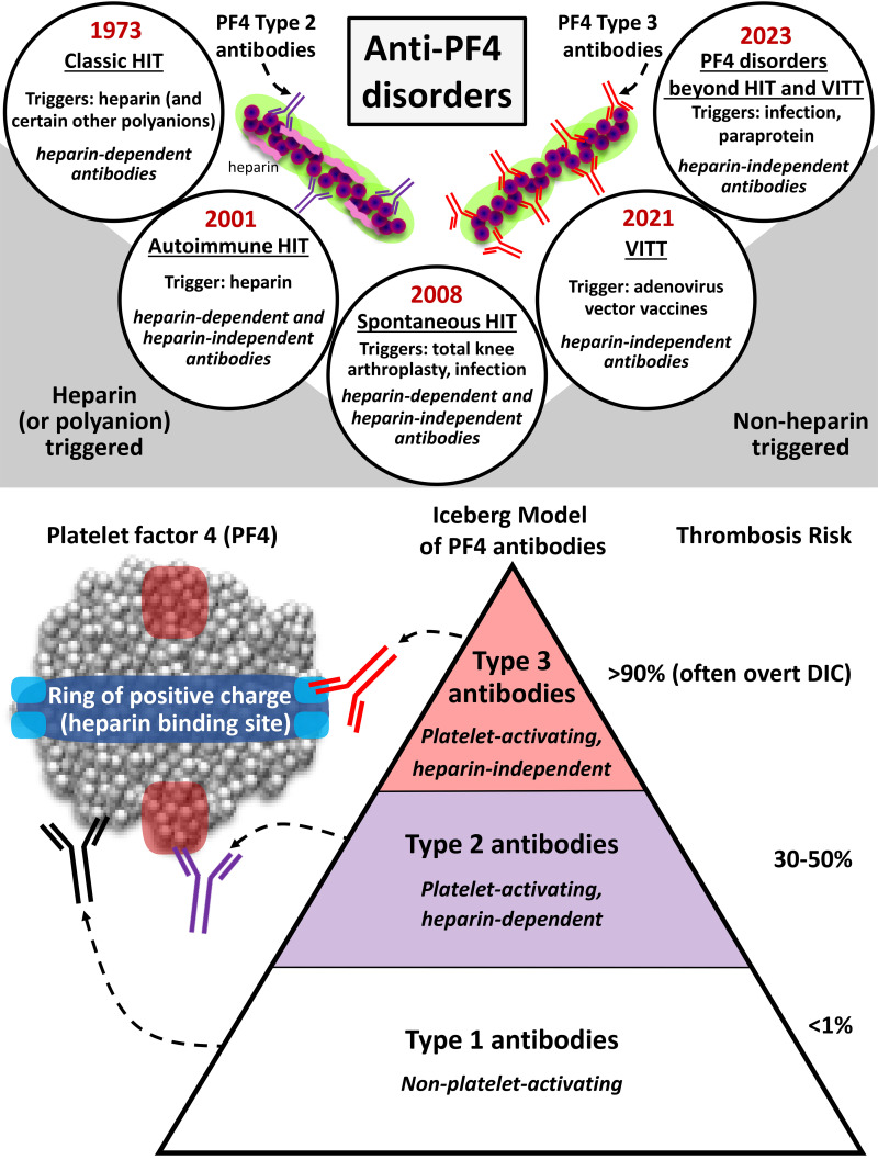
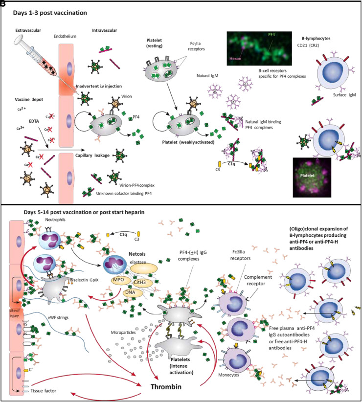
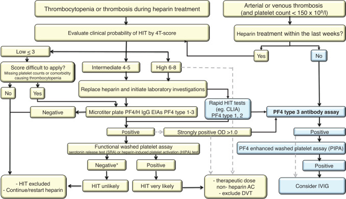
Antibodies against the chemokine platelet factor 4 (PF4) occur often, but only those that activate platelets induce severe prothrombotic disorders with associated thrombocytopenia. Heparin-induced thrombocytopenia (HIT) is the prototypic anti-PF4 disorder, mediated by strong activation of platelets through their FcγIIa (immunoglobulin G [IgG]) receptors (FcγRIIa). Concomitant pancellular activation (monocytes, neutrophils, endothelium) triggers thromboinflammation with a high risk for venous and arterial thrombosis. The classic concept of HIT is that anti-PF4/heparin IgG, recognizing antigen sites on (cationic) PF4 that form in the presence of (anionic) heparin, constitute the heparin-dependent antibodies that cause HIT. Accordingly, HIT is managed by anticoagulation with a nonheparin anticoagulant. In 2021, adenovirus vector COVID-19 vaccines triggered the rare adverse effect "vaccine-induced immune thrombotic thrombocytopenia" (VITT), also caused by anti-PF4 IgG. VITT is a predominantly heparin-independent platelet-activating disorder that requires both therapeutic-dose anticoagulation and inhibition of FcγRIIa-mediated platelet activation by high-dose intravenous immunoglobulin (IVIG). HIT and VITT antibodies bind to different epitopes on PF4; new immunoassays can differentiate between these distinct HIT-like and VITT-like antibodies. These studies indicate that (1) severe, atypical presentations of HIT ("autoimmune HIT") are associated with both HIT-like (heparin-dependent) and VITT-like (heparin-independent) anti-PF4 antibodies; (2) in some patients with severe acute (and sometimes chronic, recurrent) thrombosis, VITT-like antibodies can be identified independent of proximate heparin exposure or vaccination. We propose to classify anti-PF4 antibodies as type 1 (nonpathogenic, non- platelet activating), type 2 (heparin dependent, platelet activating), and type 3 (heparin independent, platelet activating). A key concept is that type 3 antibodies (autoimmune HIT, VITT) require anticoagulation plus an adjunct treatment, namely high-dose IVIG, to deescalate the severe anti-PF4 IgG-mediated hypercoagulability state.


Conflict of interest statement

Andreas Greinacher: grants and nonfinancial support: Aspen, Boehringer Ingelheim, MSD, Bristol Myers Squibb, Paringenix, Bayer Healthcare, Gore Inc., Rovi, Sagent, Biomarin/Prosensa, Portola, Ergomed, GTH e.V.; personal fees: Aspen, Boehringer Ingelheim, MSD, Macopharma, Bristol Myers Squibb, Chromatec, Werfen (Instrumentation Laboratory).

Theodore E. Warkentin: honoraria: Alexion, Werfen (Instrumentation Laboratory); royalties: Informa (Taylor & Francis); consultancy: Aspen Canada, Aspen Global, CSL Behring, Ergomed, Paradigm Pharmaceuticals, Octapharma, Veralox Therapeutics; research funding: Werfen (Instrumentation Laboratory).

Figures

None
Graphical abstract
Figure 1.
Figure 1.
Current concepts of the pathogenesis of HIT and VITT. The schematic presentation shown in panel B is speculative and in large part inferred from experiments in HIT. The schematic presentation of the downstream prothrombotic process shown in panel C is largely substantiated by experimental data, some performed with VITT antibodies, others with HIT antibodies. (A) After heparin exposure, positively charged PF4 binds to negatively charged heparin; PF4/heparin complexes are formed. Natural IgM binds to the complexes, and complement factor C3 binds to the natural IgM. Complexes of PF4/heparin, natural IgM, and C3 bind to B cells via the complement receptor CR2 (CD21). B cells expressing the receptor for the HIT antigen on PF4 also bind to the PF4/heparin complexes, thus activating B cells to produce anti-PF4/heparin antibodies. The far left of the panel shows atomic force microscopy images of PF4 alone (upper) and formation of PF4/heparin clusters when PF4 is incubated with heparin (lower). (B) After vaccination, PF4 comes in contact with vaccine constituents and activates B cells. Left: It has been proposed that a direct inadvertent breach in the microvasculature at the vaccination site by IV injection or by disruption of VE-cadherin tight junctions by EDTA in ChAdOx1 nCoV allows vaccine constituents to enter the circulation. Within the circulation, adenovirus particles can bind to platelets and can also bind PF4 released by activated platelets or from the microvascular endothelium. Platelets may become activated (i) by vessel injury caused by injection of vaccine, (ii) after binding of the virions to the cell surface, or (iii) by immune complexes formed between contaminating host cell line proteins in the vaccine and natural IgG antibodies against these proteins. Whether the virions themselves or another yet unknown constituent in the vaccine causes the conformational change in PF4 is unknown. Middle: Once complexes with PF4 have formed, natural IgM antibodies activate complement (as it has been shown for PF4/heparin complexes), which enhances their proximity to B-cell receptors. In a mouse model, upon IV injection of ChAdOx1, platelet-bound adenoviral particles are transported to the marginal zone of the spleen, where B cells are activated upon direct contact. However, electron microscopy and superresolution microscopy revealed complexes between PF4 and anti-PF4 VITT antibodies with amorphous constituents of the vaccine rather than virus particles. Beside the virions, other potential partners for PF4 include unassembled hexons and host cell-line proteins. However, there is little overlap in the proteins contaminating ChAdOx1 and Ad26.COV2 vaccines, which both induce anti-PF4 VITT antibodies. Right: Eventually, complexes of PF4 and vaccine come in contact with B cells, expressing a cognate Ig receptor for PF4, either as fluid-phase complexes, as virion-PF4 complexes, or as complexes presented by platelets. (C) From right to left: After clonal expansion and isotype switching of one or a few B-cell clones in VITT, or polyclonal B cells in HIT, high-titer IgG anti-PF4 antibodies are released into the circulation. Immune complexes containing PF4 (or PF4/heparin complexes) and anti-PF4 IgG cluster and signal through FcRγIIA, which generates procoagulant platelets, induces platelet/neutrophil aggregates, and stimulates NETosis by neutrophils. DNA released by NETosis amplifies immune injury and activates complement, which deposits on the endothelium. Endothelial cells become activated, expressing tissue factor and releasing von Willebrand factor (VWF). VWF binds PF4 and subsequently anti-PF4 antibodies, which in turn further activates neutrophils and further propagates thrombin generation. To reduce complexity, multimolecular PF4 complexes in VITT are shown and not the multimolecular PF4/heparin complexes in HIT as shown in panel A. HS, heparan sulfate; MPO, myeloperoxidase. Data modified from Cines and Greinacher.
Figure 1.
Figure 1.
Current concepts of the pathogenesis of HIT and VITT. The schematic presentation shown in panel B is speculative and in large part inferred from experiments in HIT. The schematic presentation of the downstream prothrombotic process shown in panel C is largely substantiated by experimental data, some performed with VITT antibodies, others with HIT antibodies. (A) After heparin exposure, positively charged PF4 binds to negatively charged heparin; PF4/heparin complexes are formed. Natural IgM binds to the complexes, and complement factor C3 binds to the natural IgM. Complexes of PF4/heparin, natural IgM, and C3 bind to B cells via the complement receptor CR2 (CD21). B cells expressing the receptor for the HIT antigen on PF4 also bind to the PF4/heparin complexes, thus activating B cells to produce anti-PF4/heparin antibodies. The far left of the panel shows atomic force microscopy images of PF4 alone (upper) and formation of PF4/heparin clusters when PF4 is incubated with heparin (lower). (B) After vaccination, PF4 comes in contact with vaccine constituents and activates B cells. Left: It has been proposed that a direct inadvertent breach in the microvasculature at the vaccination site by IV injection or by disruption of VE-cadherin tight junctions by EDTA in ChAdOx1 nCoV allows vaccine constituents to enter the circulation. Within the circulation, adenovirus particles can bind to platelets and can also bind PF4 released by activated platelets or from the microvascular endothelium. Platelets may become activated (i) by vessel injury caused by injection of vaccine, (ii) after binding of the virions to the cell surface, or (iii) by immune complexes formed between contaminating host cell line proteins in the vaccine and natural IgG antibodies against these proteins. Whether the virions themselves or another yet unknown constituent in the vaccine causes the conformational change in PF4 is unknown. Middle: Once complexes with PF4 have formed, natural IgM antibodies activate complement (as it has been shown for PF4/heparin complexes), which enhances their proximity to B-cell receptors. In a mouse model, upon IV injection of ChAdOx1, platelet-bound adenoviral particles are transported to the marginal zone of the spleen, where B cells are activated upon direct contact. However, electron microscopy and superresolution microscopy revealed complexes between PF4 and anti-PF4 VITT antibodies with amorphous constituents of the vaccine rather than virus particles. Beside the virions, other potential partners for PF4 include unassembled hexons and host cell-line proteins. However, there is little overlap in the proteins contaminating ChAdOx1 and Ad26.COV2 vaccines, which both induce anti-PF4 VITT antibodies. Right: Eventually, complexes of PF4 and vaccine come in contact with B cells, expressing a cognate Ig receptor for PF4, either as fluid-phase complexes, as virion-PF4 complexes, or as complexes presented by platelets. (C) From right to left: After clonal expansion and isotype switching of one or a few B-cell clones in VITT, or polyclonal B cells in HIT, high-titer IgG anti-PF4 antibodies are released into the circulation. Immune complexes containing PF4 (or PF4/heparin complexes) and anti-PF4 IgG cluster and signal through FcRγIIA, which generates procoagulant platelets, induces platelet/neutrophil aggregates, and stimulates NETosis by neutrophils. DNA released by NETosis amplifies immune injury and activates complement, which deposits on the endothelium. Endothelial cells become activated, expressing tissue factor and releasing von Willebrand factor (VWF). VWF binds PF4 and subsequently anti-PF4 antibodies, which in turn further activates neutrophils and further propagates thrombin generation. To reduce complexity, multimolecular PF4 complexes in VITT are shown and not the multimolecular PF4/heparin complexes in HIT as shown in panel A. HS, heparan sulfate; MPO, myeloperoxidase. Data modified from Cines and Greinacher.
Figure 2.
Figure 2.
Flow chart for the diagnosis of anti–PF4-antibody-induced prothrombotic disorders. Anti–PF4 antibody-mediated disorders should be clinically diagnosed and laboratory testing only performed upon reasonable clinically suspicion. The yellow part of the figure presents the current diagnostic flow for heparin-induced thrombocytopenia. Anti-PF4/heparin antibody antigen tests are differentiated between microtiter plate-based EIAs and rapid HIT tests (Table 2). The light-blue part of the figure shows the workflow when clinically applicable immunoassays for anti-PF4 type 3 antibodies are available (currently under development). As the blue section of the workflow is untested, the decision to test for anti-PF4 antibodies should include consideration of the pretest probability: if there are other obvious reasons to explain thrombosis and thrombocytopenia, such as cancer or intensive care unit treatment, testing is probably not indicated. The dotted lines show the workflow based on clinical considerations, bypassing some diagnostics steps; for example, a typical presentation of HIT with a high 4Ts score of 6, 7, or 8 points should immediately prompt therapeutic-dose alternative anticoagulation, and laboratory test results are used to confirm the diagnosis. *Functional assays can be more sensitive if PF4 is added. Clinically nonrelevant anti-PF4 type 1 antibodies (detected in up to 20% of heparin-treated patients and up to 8% of COVID-19 vaccinated individuals) do not require treatment change. AC, anticoagulation; CLIA, chemiluminescence-based immunoassay; DVT, deep vein thrombosis; OD, optical density.

Similar articles

Cited by

References

    1. Warkentin TE, Greinacher A.. Spontaneous HIT syndrome: knee replacement, infection, and parallels with vaccine-induced immune thrombotic thrombocytopenia. Thromb Res. 2021;204(August):40-51. doi:10.1016/j.thromres.2021.05.018. - DOI - PubMed
    1. Wagner DD, Heger LA. Thromboinflammation: from atherosclerosis to COVID-19. Arterioscler Thromb Vasc Biol. 2022;42(9):1103-1112. doi:10.1161/ATVBAHA.122.317162. - DOI - PMC - PubMed
    1. Warkentin TE. Heparin-induced thrombocytopenia-associated thrombosis: from arterial to venous to venous limb gangrene. J Thromb Haemost. 2018;16(11):2128-2132. doi:10.1111/jth.14264. - DOI - PubMed
    1. Cuker A, Arepally GM, Chong BH, et al.. American Society of Hematology 2018 guidelines for management of venous thromboembolism: heparin-induced thrombocytopenia. Blood Adv. 2018;2(22):3360-3392. doi:10.1182/bloodadvances.2018024489. - DOI - PMC - PubMed
    1. Warkentin TE. High-dose intravenous immunoglobulin for the treatment and prevention of heparin-induced thrombocytopenia: a review. Expert Rev Hematol. 2019;12(8):685-698. doi:10.1080/17474086.2019.1636645. - DOI - PubMed

MeSH terms