. 2023 Nov 21;12(23):2668.
doi: 10.3390/cells12232668.

Patterns of Gene Expression, Splicing, and Allele-Specific Expression Vary among Macular Tissues and Clinical Stages of Age-Related Macular Degeneration

Affiliations

Patterns of Gene Expression, Splicing, and Allele-Specific Expression Vary among Macular Tissues and Clinical Stages of Age-Related Macular Degeneration

Treefa Shwani et al. Cells. .

Abstract

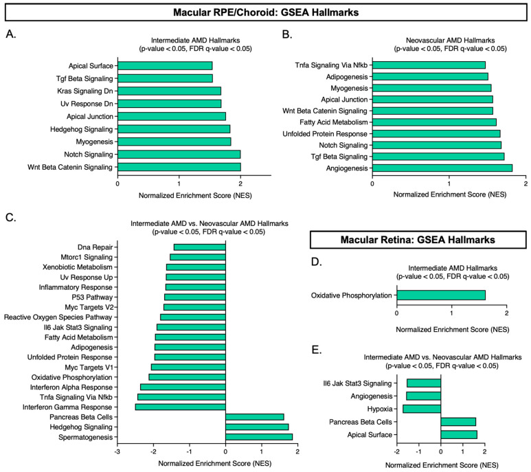
Age-related macular degeneration (AMD) is a leading cause of blindness, and elucidating its underlying disease mechanisms is vital to the development of appropriate therapeutics. We identified differentially expressed genes (DEGs) and differentially spliced genes (DSGs) across the clinical stages of AMD in disease-affected tissue, the macular retina pigment epithelium (RPE)/choroid and the macular neural retina within the same eye. We utilized 27 deeply phenotyped donor eyes (recovered within a 6 h postmortem interval time) from Caucasian donors (60-94 years) using a standardized published protocol. Significant findings were then validated in an independent set of well-characterized donor eyes (n = 85). There was limited overlap between DEGs and DSGs, suggesting distinct mechanisms at play in AMD pathophysiology. A greater number of previously reported AMD loci overlapped with DSGs compared to DEGs between disease states, and no DEG overlap with previously reported loci was found in the macular retina between disease states. Additionally, we explored allele-specific expression (ASE) in coding regions of previously reported AMD risk loci, uncovering a significant imbalance in C3 rs2230199 and CFH rs1061170 in the macular RPE/choroid for normal eyes and intermediate AMD (iAMD), and for CFH rs1061147 in the macular RPE/choroid for normal eyes and iAMD, and separately neovascular AMD (NEO). Only significant DEGs/DSGs from the macular RPE/choroid were found to overlap between disease states. STAT1, validated between the iAMD vs. normal comparison, and AGTPBP1, BBS5, CERKL, FGFBP2, KIFC3, RORα, and ZNF292, validated between the NEO vs. normal comparison, revealed an intricate regulatory network with transcription factors and miRNAs identifying potential upstream and downstream regulators. Findings regarding the complement genes C3 and CFH suggest that coding variants at these loci may influence AMD development via an imbalance of gene expression in a tissue-specific manner. Our study provides crucial insights into the multifaceted genomic underpinnings of AMD (i.e., tissue-specific gene expression changes, potential splice variation, and allelic imbalance), which may open new avenues for AMD diagnostics and therapies specific to iAMD and NEO.

Keywords: age-related macular degeneration (AMD); allele-specific expression (ASE); and AMD therapies; intermediate AMD (iAMD); macular retina; macular retinal pigment epithelium (RPE)/choroid; microRNAs (miRNAs); neovascular AMD (NEO); splicing; tissue-specific gene expression.


Conflict of interest statement

The authors declare no conflict of interest.

Figures

Figure 1
Figure 1
Violin plot of Log10-transformed FPKM counts from 27 donor eye samples with both the macular RPE/choroid and macular retina shown. Abbreviations: AMD, age-related macular degeneration, RPE, retinal pigment epithelium, FPKM, fragments per kilobase of transcript per million mapped reads.
Figure 2
Figure 2
Volcano plots of differentially expressed genes across disease states. (AF) Each dot represents one of the 26,650 genes expressed. Blue and red represent significant genes, with red indicating upregulation and blue indicating downregulation in each disease comparison. Grey dots represent genes that did not meet the significance threshold of padj < 0.05 and a fold change ≥ |1.5|. The ten most significant genes in each disease comparison are labeled. Abbreviations: AMD, age-related macular degeneration, RPE, retinal pigment epithelium.
Figure 3
Figure 3
Overlap of differentially expressed genes (DEGs) and differentially spliced genes (DSGs) between intermediate AMD (iAMD) vs. normal and neovascular AMD (NEO) vs. normal. (AD) Each circle represents the number of significant DEGs or DSGs in macular RPE (retinal pigment epithelium)/choroid and macular retina. The overlap between these two circles shows the number of overlapping genes that were regulated in the same direction between each comparison. Abbreviations: AMD, age-related macular degeneration, RPE, retinal pigment epithelium.
Figure 4
Figure 4
Volcano plots of differentially spliced genes across disease states. (AF) Each dot represents one of the 26,650 genes expressed. Blue and red represent significant genes, with red representing upregulation and blue representing downregulation in each disease comparison. Grey dots represent genes that did not meet the significance threshold of padj < 0.05 and a fold change ≥ |1.5|. The ten most significant genes in each disease comparison are labelled. Abbreviations: AMD, age-related macular degeneration, RPE, retinal pigment epithelium.
Figure 5
Figure 5
Gene set enrichment analysis (GSEA) using our normalized expression dataset. Thresholds were set based on the nominal p-value < 0.05 and FDR q-value ≤ 0.05 generated by the GSEA software, v.4.3.2. (AC) show the significant hallmarks (p-value < 0.05, FDR q-value ≤ 0.05) identified in the macular RPE/choroid across disease state. (D,E) show the significant hallmarks (p-value < 0.05, FDR q-value ≤ 0.05) identified in the macular retina across disease state. Please note: no hallmark was identified to be significantly upregulated in neovascular AMD for the macular retina. Abbreviations: GSEA, gene set enrichment analysis, AMD, age-related macular degeneration, RPE, retinal pigment epithelium.
Figure 6
Figure 6
Visualization of significant DEGs and DSGs (previously associated with AMD) found in normal macular RPE/choroid vs. normal macular retina to illustrate directionality in normal tissues. Abbreviations: AMD, age-related macular degeneration, RPE, retinal pigment epithelium, DEG, differentially expressed gene, DSG, differentially spliced gene.
Figure 7
Figure 7
Ingenuity Pathway Analysis (IPA)-generated network of our validated genes (DEGs, DSGs, and confirmed in an independent bulk RNAseq dataset): (A,B) show the 7 identified genes from NEO vs. normal; (C,D) show our 7 identified genes from NEO vs. normal combined with STAT1, our validated gene from iAMD vs. normal. Each network is overlaid with expression values/fold changes from either our DEG or DSG dataset for that disease state comparison. Red or green indicates the gene was found in our dataset and associated with increased or decreased measurement, respectively. Orange or blue indicates the gene was not in our dataset but is predicted to be associated with activation or inhibition. Further clarification is provided in the legend. Abbreviations: DEG, differentially expressed gene, DSG, differentially spliced gene.

References

    1. Ung C., Lains I., Miller J.W., Kim I.K. Current Management of Age-Related Macular Degeneration. In: Chew E.Y., Swaroop A., editors. Age-Related Macular Degeneration. Springer International Publishing; Cham, Switzerland: 2021. pp. 295–314. - PubMed
    1. Chaudhuri M., Hassan Y., Vemana P.P.S.B., Pattanashetty M.S.B., Abdin Z.U., Siddiqui H.F. Age-Related Macular Degeneration: An Exponentially Emerging Imminent Threat of Visual Impairment and Irreversible Blindness. Cureus. 2023;15:e39624. doi: 10.7759/cureus.39624. - DOI - PMC - PubMed
    1. Khachigian L.M., Liew G., Teo K.Y.C., Wong T.Y., Mitchell P. Emerging therapeutic strategies for unmet need in neovascular age-related macular degeneration. J. Transl. Med. 2023;21:133. doi: 10.1186/s12967-023-03937-7. - DOI - PMC - PubMed
    1. Tan C.S., Ngo W.K., Chay I.W., Ting D.S., Sadda S.R. Neovascular Age-Related Macular Degeneration (nAMD): A Review of Emerging Treatment Options. Clin. Ophthalmol. 2022;16:917–933. doi: 10.2147/OPTH.S231913. - DOI - PMC - PubMed
    1. Reiter G.S., Schmidt-Erfurth U. Quantitative assessment of retinal fluid in neovascular age-related macular degeneration under anti-VEGF therapy. Ther. Adv. Ophthalmol. 2022;14:25158414221083363. doi: 10.1177/25158414221083363. - DOI - PMC - PubMed

Publication types