Review
. 2023 Nov 22;12(23):2684.
doi: 10.3390/cells12232684.

Chemistry of Hydrogen Sulfide-Pathological and Physiological Functions in Mammalian Cells

Affiliations
Review

Chemistry of Hydrogen Sulfide-Pathological and Physiological Functions in Mammalian Cells

Celia María Curieses Andrés et al. Cells. .

Abstract

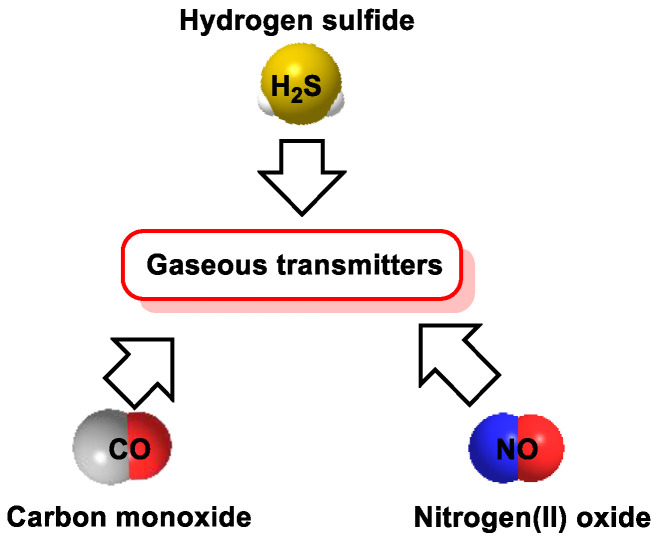
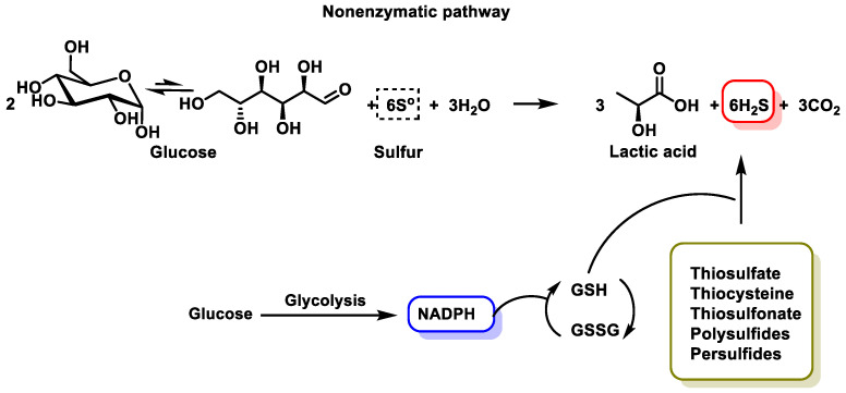
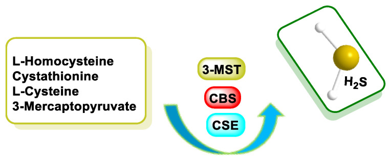
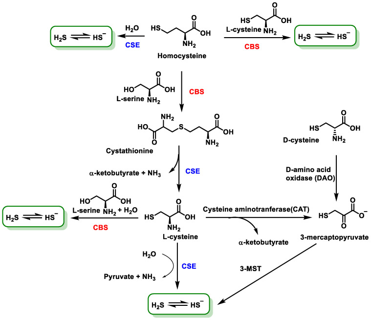
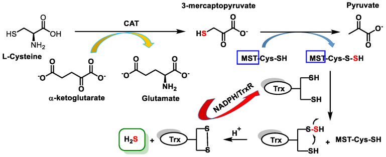
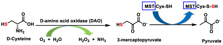
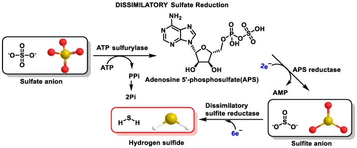
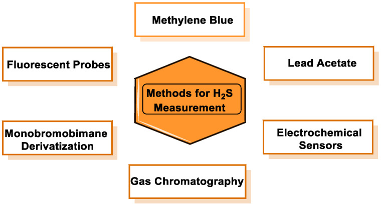
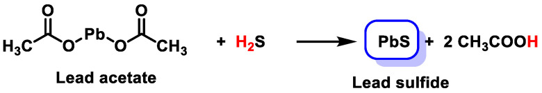
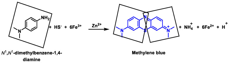
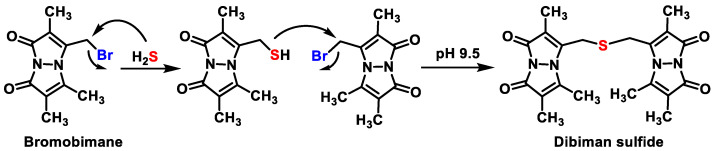
Hydrogen sulfide (H2S) was recognized as a gaseous signaling molecule, similar to nitric oxide (-NO) and carbon monoxide (CO). The aim of this review is to provide an overview of the formation of hydrogen sulfide (H2S) in the human body. H2S is synthesized by enzymatic processes involving cysteine and several enzymes, including cystathionine-β-synthase (CBS), cystathionine-γ-lyase (CSE), cysteine aminotransferase (CAT), 3-mercaptopyruvate sulfurtransferase (3MST) and D-amino acid oxidase (DAO). The physiological and pathological effects of hydrogen sulfide (H2S) on various systems in the human body have led to extensive research efforts to develop appropriate methods to deliver H2S under conditions that mimic physiological settings and respond to various stimuli. These functions span a wide spectrum, ranging from effects on the endocrine system and cellular lifespan to protection of liver and kidney function. The exact physiological and hazardous thresholds of hydrogen sulfide (H2S) in the human body are currently not well understood and need to be researched in depth. This article provides an overview of the physiological significance of H2S in the human body. It highlights the various sources of H2S production in different situations and examines existing techniques for detecting this gas.

Keywords: chemistry; gasotransmitter; hydrogen sulfide; physiology.


Conflict of interest statement

Eduardo Perez Lebeña is an employee of Sistemas de Biotecnología y Recursos Naturales. The other authors declare no conflict of interest. The funders had no role in the design of the study; in the collection, analyses or interpretation of data; in the writing of the manuscript; or in the decision to publish the results.

Figures

Figure 1
Figure 1
NO, CO and H2S are gaseous signaling molecules involved in biological functions.
Figure 2
Figure 2
H2S molecule structural formula, molecular geometry, angle and Lewis structure.
Figure 3
Figure 3
Various non-enzymatic routes of H2S synthesis. In the presence of reducing equivalents such as NADPH and NADH, reactive sulfur species in persulfides, thiosulfate and polysulfides are reduced into H2S and other metabolites. GSH is glutathione and GSSG is glutathione disulfide.
Figure 4
Figure 4
The biosynthesis of H2S mammalian cells primarily involves cystathionine β-synthase (CBS), cystathionine-γ-lyase (CSE) and 3-mercaptopyruvate sulfurtransferase (3MST).
Figure 5
Figure 5
Main route of H2S synthesis mediated by the enzymes CBS, CSE and 3MST.
Figure 6
Figure 6
H2S synthesis via the 3MST/CAT pathway.
Figure 7
Figure 7
Formation of 3-mercaptopyruvate from D-cysteine.
Figure 8
Figure 8
Dissimilatory sulfate reduction pathway.
Figure 9
Figure 9
Mineral-acid-catalyzed H2S synthesis.
Figure 10
Figure 10
The metabolic processes involved in the breakdown and utilization of H2S. H2S is degraded by various enzymatic reactions. The enzymes rhodanese, bisulfide methyltransferase (BMT) and thiosulfate reductase (TSR) play critical roles in catalyzing the conversion of H2S to thiocyanate, methanethiol and thiosulfate, respectively. Oxidation of thiosulfate to sulfite can occur through the enzymatic action of thiosulfate sulfurtransferase (TSST), followed by further oxidation to sulfate. H2S reacts with hemoglobin, resulting in the formation of sulfohemoglobin. In addition, H2S combines with proteins present in the tissue, resulting in the formation of a bound sulfur pool.
Figure 11
Figure 11
The two alternative modes for sulfide oxidation.
Figure 12
Figure 12
Reaction mechanism postulated for the oxidation of H2S by MetHb. Methemoglobin binds to H2S and oxidizes it to a mixture of thiosulfate and hydropolysulfides.
Figure 13
Figure 13
HS reaction with two different nucleophiles.
Figure 14
Figure 14
H2S dissociation equilibria.
Figure 15
Figure 15
Reaction of H2S with oxygen.
Figure 16
Figure 16
Chemical reactivity of H2S.
Figure 17
Figure 17
Reaction of disulfide reduction by HS.
Figure 18
Figure 18
Examples of diseases related to low levels of H2S.
Figure 19
Figure 19
Physiological roles of H2S.
Figure 20
Figure 20
H2S signaling mechanisms.
Figure 21
Figure 21
KATP channel subunits.
Figure 22
Figure 22
Oxidation of H2S by H2O2, ONOOH and HOCl. Formation of sulfenic acid (HSOH).
Figure 23
Figure 23
Oxidation of H2S and formation of the sulfiyl radical (HS/S•−).
Figure 24
Figure 24
Chain reactions produced by the sulfiyl radical (HS/S•−), which is an oxidant.
Figure 25
Figure 25
Reaction of H2S with peroxynitrite with formation of sulfinyl nitrite.
Figure 26
Figure 26
Mechanisms of protein persulfidation.
Figure 27
Figure 27
Hydropersulfide cyanolysis reaction.
Figure 28
Figure 28
Persulfide detection reaction.
Figure 29
Figure 29
Classical iodometric titration.
Figure 30
Figure 30
Methods for H2S measurement.
Figure 31
Figure 31
Hydrogen sulfide reacts with lead acetate to form a brown solid of lead sulfide (PbS).
Figure 32
Figure 32
Reaction in the methylene blue method for sulfide detection.
Figure 33
Figure 33
Bromobrimane reaction to obtain dibiman sulfide.
Figure 34
Figure 34
Fluorescent probes for the detection of H2S using the reduction of azide (ac) or nitro (d) groups.
Figure 35
Figure 35
Reaction of MitoA with H2S to form MitoN.
Figure 36
Figure 36
Structures of ratiometric H2S probes that function by disrupting the conjugated p-system within a fluorophore. Also shown is the process by which these probes react with H2S.
Figure 37
Figure 37
Fluorescent probe for the detection of hydrogen sulfide on the basis of H2S-mediated benzodithiolone formation.
Figure 38
Figure 38
Fluorescent probes and reaction of methyl (E)-3-(5-(3-(3,5-difluorophenyl)-1-phenyl-1H-pyrazol-5-yl)-2-formylphenyl)acrylate with H2S.
Figure 39
Figure 39
Use of azamacrocyclic copper(II) ion complex chemistry to modulate fluorescence in the fluorescent probe for the detection of H2S, known as HSip-1.
Figure 40
Figure 40
Sodium acid sulfide and sodium sulfide spontaneously release H2S.
Figure 41
Figure 41
The biological functions of allicin and its secondary metabolites.
Figure 42
Figure 42
Enzymatic conversion of allin to allicin. Two molecules of 2-propenesulfenic acid interact to form allicin and remove water.
Figure 43
Figure 43
Organosulfur compounds derived from allicin.
Figure 44
Figure 44
The main compounds found in intact garlic cloves.
Figure 45
Figure 45
Proposed mechanism of H2S release from trisulfide in the presence of GSH by initial GSH attack on sulfur and a second GSH attack on α-carbon with the formation of S-allyi glutathione.
Figure 46
Figure 46
Natural sulfur compounds present in plants that have been associated with the formation of hydrogen sulfide.
Figure 47
Figure 47
Organic H2S donors and the mechanisms by which they produce H2S.
Figure 48
Figure 48
Chemical synthesis of ZYZ-803.
Figure 49
Figure 49
Mitochondrial H2S donors.
Figure 50
Figure 50
H2S release mechanism by AP39 and H2S release mechanism by AP123.
Figure 51
Figure 51
Reaction mechanism of thiol-dependent H2S release by MitoPerSulf. Reactions with GSH are shown.
Figure 52
Figure 52
Examples of approved and used drugs containing sulfur-containing functional groups or residues and therapeutic applications.
Figure 53
Figure 53
Structural formula of 2-acetoxybenzoic acid and 4-(3-thioxo-3H-1,2-dithiol-5-yl) phenyl-2-acetoxybenzoate.
Figure 54
Figure 54
The structure of the H2S-releasing agents derived from l-DOPA: ACS83, ACS84, ACS85 and ACS86.

Similar articles

Cited by

References

    1. Mustafa A.K., Gadalla M.M., Sen N., Kim S., Mu W., Gazi S.K., Barrow R.K., Yang G., Wang R., Snyder S.H. H2S signals through protein S-sulfhydration. Sci. Signal. 2009;2:ra72. doi: 10.1126/scisignal.2000464. - DOI - PMC - PubMed
    1. Mathai J.C., Missner A., Kügler P., Saparov S.M., Zeidel M.L., Lee J.K., Pohl P. No facilitator required for membrane transport of hydrogen sulfide. Proc. Natl. Acad. Sci. USA. 2009;106:16633–16638. doi: 10.1073/pnas.0902952106. - DOI - PMC - PubMed
    1. Whiteman M., Winyard P.G. Hydrogen sulfide and inflammation: The good, the bad, the ugly and the promising. Expert. Rev. Clin. Pharmacol. 2011;4:13–32. doi: 10.1586/ecp.10.134. - DOI - PubMed
    1. Kimura H., Shibuya N., Kimura Y. Hydrogen sulfide is a signaling molecule and a cytoprotectant. Antioxid. Redox Signal. 2012;17:45–57. doi: 10.1089/ars.2011.4345. - DOI - PMC - PubMed
    1. Szabó C. Hydrogen sulphide and its therapeutic potential. Nat. Rev. Drug Discov. 2007;6:917–935. doi: 10.1038/nrd2425. - DOI - PubMed

Publication types

LinkOut - more resources