. 2023 Nov 23;12(23):2694.
doi: 10.3390/cells12232694.

Calreticulin Regulates SARS-CoV-2 Spike Protein Turnover and Modulates SARS-CoV-2 Infectivity

Affiliations

Calreticulin Regulates SARS-CoV-2 Spike Protein Turnover and Modulates SARS-CoV-2 Infectivity

Nader Rahimi et al. Cells. .

Abstract

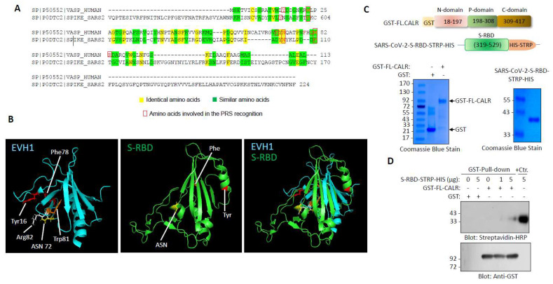
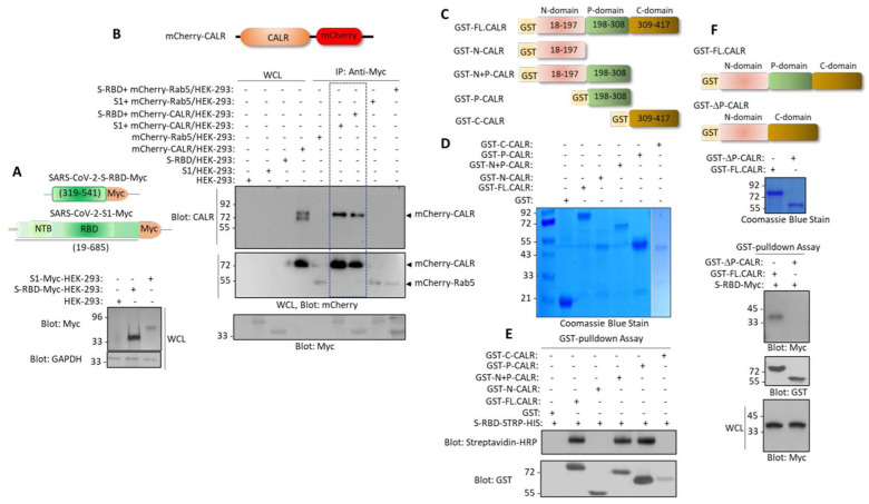
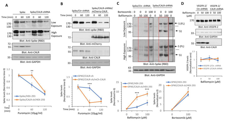
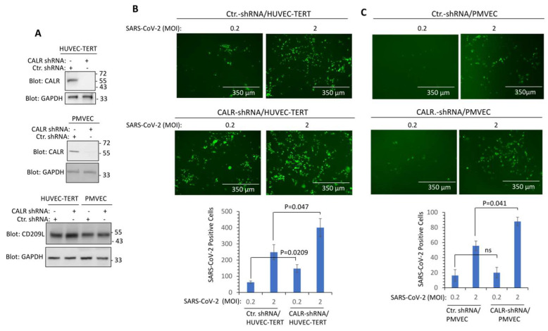
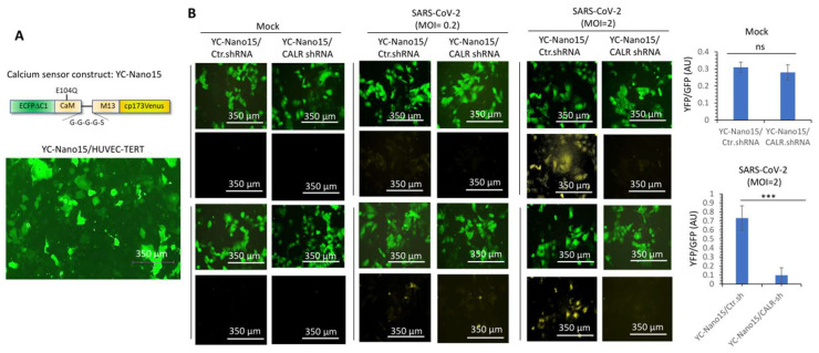
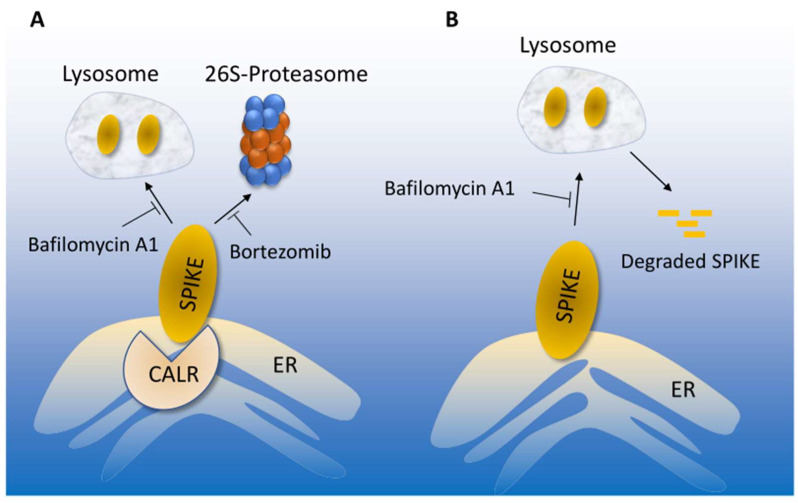
Cardiovascular complications are major clinical hallmarks of acute and post-acute coronavirus disease 2019 (COVID-19). However, the mechanistic details of SARS-CoV-2 infectivity of endothelial cells remain largely unknown. Here, we demonstrate that the receptor binding domain (RBD) of the SARS-CoV-2 spike (S) protein shares a similarity with the proline-rich binding ena/VASP homology (EVH1) domain and identified the endoplasmic reticulum (ER) resident calreticulin (CALR) as an S-RBD interacting protein. Our biochemical analysis showed that CALR, via its proline-rich (P) domain, interacts with S-RBD and modulates proteostasis of the S protein. Treatment of cells with the proteasomal inhibitor bortezomib increased the expression of the S protein independent of CALR, whereas the lysosomal/autophagy inhibitor bafilomycin 1A, which interferes with the acidification of lysosome, selectively augmented the S protein levels in a CALR-dependent manner. More importantly, the shRNA-mediated knockdown of CALR increased SARS-CoV-2 infection and impaired calcium homeostasis of human endothelial cells. This study provides new insight into the infectivity of SARS-CoV-2, calcium hemostasis, and the role of CALR in the ER-lysosome-dependent proteolysis of the spike protein, which could be associated with cardiovascular complications in COVID-19 patients.

Keywords: COVID-19; S-RBD; SARS-CoV-2; calreticulin; endothelial cells; intracellular calcium homeostasis; spike protein.


Conflict of interest statement

The authors declare no conflict of interest.

Figures

Figure 1
Figure 1
S-RBD structure resembles EVH1 domain and interacts with CALR: (A) Amino acid alignment of EVH1 domain of human VASP protein with SARS-CoV-2 S RBD. (B) Three-dimensional structures of EVH1 and S-RBD. (C) Schematic of GST-fusion of full length CLAR and S-RBD-STRP-HIS constructs with Coomassie blue staining of GST-CALR and S-RBD-STRP-HIS (5 µg). (D) GST pull-down experiment showing the binding of a purified S-RBD-STRP-HIS protein with GST-CALR.
Figure 2
Figure 2
Spike protein interacts with the P-domain of CALR. (A) Schematic of S1-c-Myc subunit and S-RBD-c-Myc constructs and their expression in HEK-293 cells. (B) In vivo binding of S1-c-Myc and S-RBD-Myc with mCherry-CALR in HEK-293 cells. The mCherry-Rab5 construct was used as a negative control. (C) Schematics of GST-fusion CALR proteins. (D) Coomassie blue staining of GST-CALR proteins. (E) In vitro GST pull-down assay showing the binding of P-domain containing GST-CALR proteins with S-RBD-STRP-HIS. (F) Schematics of GST full-length (FL) CALR and GST-P-domain-truncated CALR (GST-ΔP-CALR), Coomassie blue staining, and the GST the pull-down assay showing that deletion of P domain abolishes the binding of CALR with S-RBD. WCL, whole cell lysate.
Figure 3
Figure 3
CALR regulates the lysosome-dependent proteolysis of the spike protein. (A) HEK-293 cells expressing spike-C9 alone or co-expressing spike with CALR-shRNA were treated with puromycin (10 µg/mL) for 0, 60, or 120 min. Cells were lysed and whole cell lysates were subjected to Western blot analysis using an anti-spike (RBD) or anti-CALR antibodies. Both short and long exposure of the blots are shown. The graph is a representative of three independent experiments. ** p < 0.01. (B) HEK-293 cells co-expressing spike with CALR-shRNA or spike-C9 with CALR-shRNA and mCherry-CALR were subjected to puromycin treatment and analyzed as described in panel A. (C) HEK-293 cells expressing spike-C9 alone or co-expressing spike with CALR-shRNA were treated with bafilomycin-1A with varying concentrations or with bortezomib (100 µM) for 12 h. Cells were lysed and whole cell lysates were subjected to Western blot analysis as shown in panel A. The graphs are representative of three independent experiments. (D) HEK-293 cells co-expressing VEGFR-2, control shRNA, or CALR-shRNA were treated with bafilomycin-1A with varying concentrations as indicated. Cells were lysed and whole cell lysates were subjected to Western blot analysis as shown in panel (A). The graph is representative of three independent experiments. Image J software (version v1.54g) was used to quantify the blots and the values of the spike protein and VEGFR-2 in each lane were normalized to the corresponding control baselines (time 0 or untreated group). * p < 0.05, ** p < 0.01 and *** p < 0.001.
Figure 4
Figure 4
Knockdown of CALR in human endothelial cells increases SARS-CoV-2 infection. (A) Western blot analysis shows the expression of CALR in the parental and lentivirus CALR-shRNA expressing endothelial cells. (B) HUVEC-TERT cells expressing control shRNA or CALR-shRNA were seeded in 96-well plates (triplicate per group). The next day, the cells were infected with SARS-CoV-2-mNG at an MOI of 0.2 or 2. Twenty-four hours post-infection, cells were fixed and analyzed by fluorescence microscopy. The graph is representative of SARS-CoV-2-mNG+ cells (triplicate well per group, two independent experiments). (C) Human primary pulmonary microvascular endothelial cells (PMVECs) expressing control shRNA or CALR-shRNA were prepared and infected with SARS-CoV-2-mNG as shown in panel (B). Graph is representative of SARS-CoV-2-mNG+ cells (triplicate well per group). ns, not statistically significant.
Figure 5
Figure 5
CALR is required for SARS-CoV-2-induced intracellular calcium release in human endothelial cells. (A) Expression of YC-Nano15 construct in HUVEC-TERT cells. (B) HUVEC-TERT cells expressing YC-Nano15 and HUVEC-TERT cells expressing CALR-shRNA with YC-Nano15 were seeded in 96-well plates (triplicate per group). The next day, the cells were infected with mock or SARS-CoV-2 at an MOI of 0.2 or 2. Twenty-four hours post-infection, the cells were fixed and analyzed by fluorescence microscopy. Two images per group are shown. GFP+ cells indicate the expression of YC-Nano15 in an inactive form. Yellow+ cells indicate the activated form of YC-Nano15. Graphs are representative of YFP/GFP ratio (triplicate well per group). ns, not statistically significant; *** p < 0.001.
Figure 6
Figure 6
Proposed model of CALR-mediated SARS-CoV-2 spike proteostasis. (A) SARS-CoV-2 spike protein degradation is regulated by lysosome and 26S-proteosome, which could be inhibited by bafilomycin or bortezomib, respectively. Interaction of the spike with CALR mediates the lysosome-dependent proteolysis of the spike protein. (B) In the absence of CALR, spike protein escapes from the lysosome-mediated degradation, which could lead to increased infection.

Similar articles

References

    1. Amraei R.W., Yin W., Napoleon M.A., Suder E.L., Berrigan J., Zhao Q., Olejnik J., Chandler K.B., Xia C., Feldman J., et al. CD209L/L-SIGN and CD209/DC-SIGN Act as Receptors for SARS-CoV-2. ACS Cent. Sci. 2021;7:1156–1165. doi: 10.1021/acscentsci.0c01537. - DOI - PMC - PubMed
    1. Amraei R., Xia C., Olejnik J., White M.R., Napoleon M.A., Lotfollahzadeh S., Hauser B.M., Schmidt A.G., Chitalia V., Muhlberger E., et al. Extracellular vimentin is an attachment factor that facilitates SARS-CoV-2 entry into human endothelial cells. Proc. Natl. Acad. Sci. USA. 2022;119:e2113874119. doi: 10.1073/pnas.2113874119. - DOI - PMC - PubMed
    1. Hoffmann M., Kleine-Weber H., Schroeder S., Krüger N., Herrler T., Erichsen S., Schiergens T.S., Herrler G., Wu N., Nitsche A., et al. SARS-CoV-2 Cell Entry Depends on ACE2 and TMPRSS2 and Is Blocked by a Clinically Proven Protease Inhibitor. Cell. 2020;181:271–280.e278. doi: 10.1016/j.cell.2020.02.052. - DOI - PMC - PubMed
    1. Rahimi N. C-type Lectin CD209L/L-SIGN and CD209/DC-SIGN: Cell Adhesion Molecules Turned to Pathogen Recognition Receptors. Biology. 2020;10:1. doi: 10.3390/biology10010001. - DOI - PMC - PubMed
    1. Lan J., Ge J., Yu J., Shan S., Zhou H., Fan S., Zhang Q., Shi X., Wang Q., Zhang L., et al. Structure of the SARS-CoV-2 spike receptor-binding domain bound to the ACE2 receptor. Nature. 2020;581:215–220. doi: 10.1038/s41586-020-2180-5. - DOI - PubMed

Publication types