. 2023 Nov 24;12(23):2697.
doi: 10.3390/cells12232697.

Cytological Study of Topical Effect of Azelastine Hydrochloride on the Nasal Mucous Membrane Cells in Various Nasal Rhinitis Types

Affiliations

Cytological Study of Topical Effect of Azelastine Hydrochloride on the Nasal Mucous Membrane Cells in Various Nasal Rhinitis Types

Ewa Trybus et al. Cells. .

Abstract

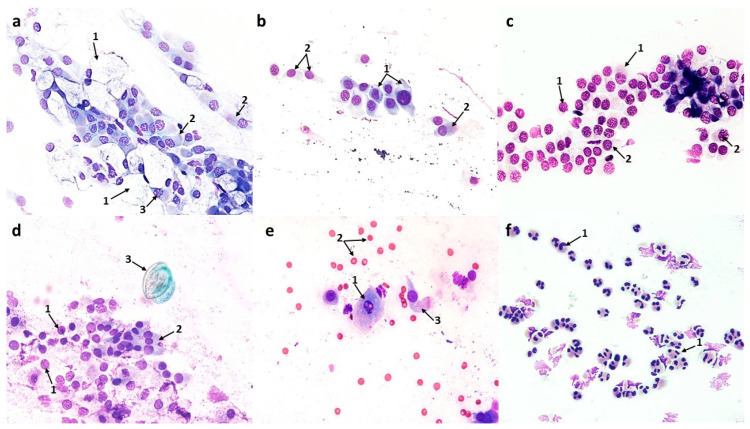
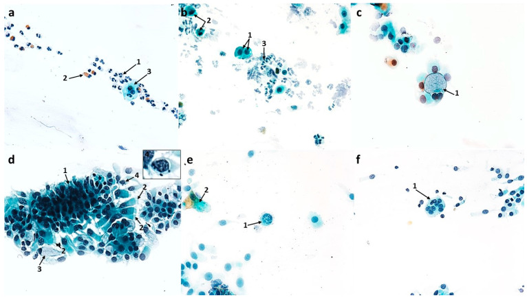
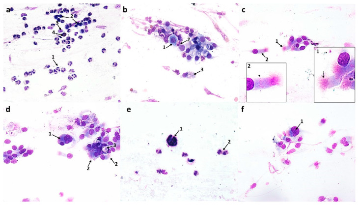
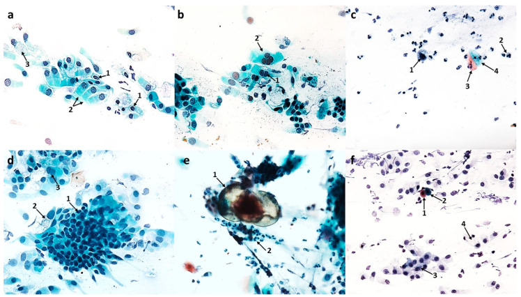
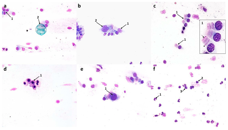
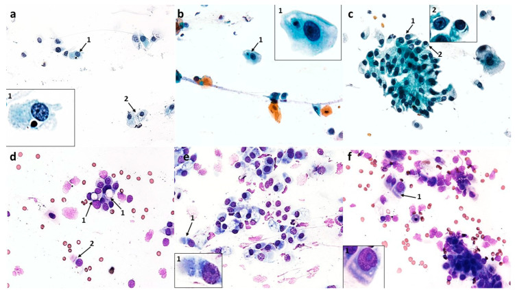
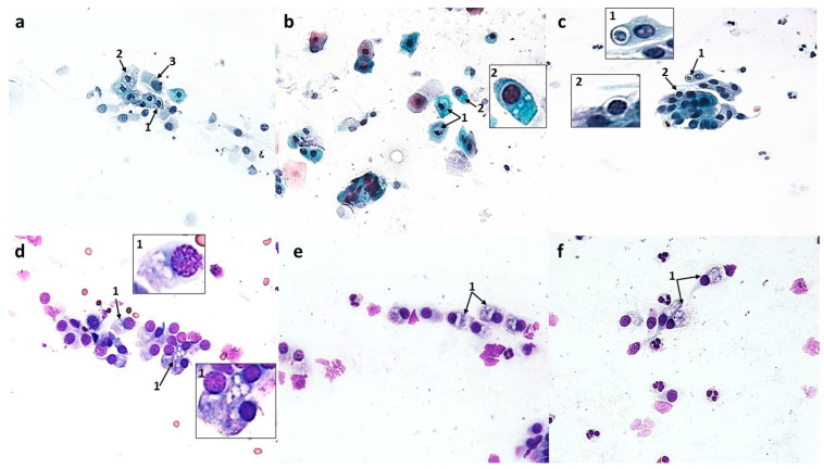
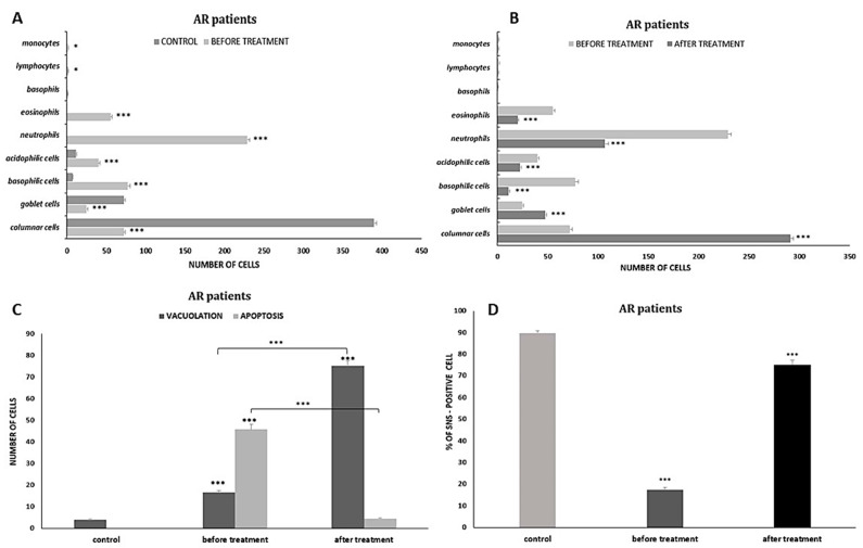
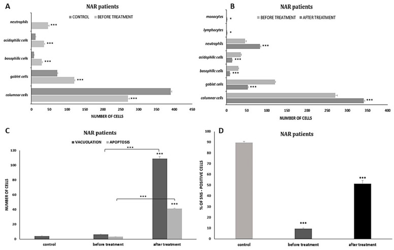
Previous reports on the benefits of using local therapy with azelastine in rhinitis focus on the assessment of clinical symptoms and the analysis of nasal lavage for the presence of inflammatory cells and the expression of adhesion molecules. Little attention has been paid to studies assessing the effect of azelastine on individual cytotypes of the nasal mucosa, especially epithelial cells, also in the context of inducing morphological changes. The aim of this study was the cytological analysis of swabs taken from the surface of the nasal mucosa of patients with allergic rhinitis (AR) and nonallergic/vasomotor rhinitis (NAR/VMR) who were subjected to 4 weeks of therapy with azelastine and then comparing the obtained results with the pre-treatment condition. The technique of obtaining materials for cytoanalysis included sampling, staining of smears, microscopic analysis, and preparation of cytograms. Our studies confirmed the therapeutic benefits of azelastine in both study groups. Significant changes were demonstrated, confirming the regeneration of ciliated cells and the induction of autophagy and apoptosis in epithelial cells. Such changes indicate new mechanisms of action of azelastine, which play a significant role in restoring homeostasis in the nasal mucosa. The presented research also results in a detailed description of cytological changes in both studied rhinitis types, which complements the knowledge regarding prognostic indicators.

Keywords: azelastine hydrochloride; nasal cytology; rhinitis.


Conflict of interest statement

The authors declare no conflict of interest.

Figures

Figure 1
Figure 1
The cytology of normal nasal epithelium under light microscopy (×400): (a) MGG staining: 1—columnar cells with normal ciliary apparatus and visible hyperchromatic supranuclear striae (SNS+ cells), 2—binucleated columnar cell, 3—columnar cell with cytoplasm vacuolization, 4—perinuclear halo in a columnar cell. (b) Papanicolaou staining: 1—columnar cells with normal ciliary apparatus and evident SNS (SNS+ cells), 2—goblet cell. (c) Distribution of nasal mucosa cells in a normal cytogram.
Figure 2
Figure 2
The cytology of the nasal epithelium of patients with allergic rhinitis (AR) before azelastine treatment under light microscopy (×400): Papanicolaou staining: (a) 1—eosinophils, 2—bacteria. (b) 1—degranulated eosinophils, 2—columnar cells with absence of ciliary apparatus (SNS cells), 3—columnar cells with rarefaction of ciliary apparatus (SNS cells). (c) 1—giant goblet cells filled with mucus, 2—columnar cells with absence of ciliary apparatus (SNS cells), 3—columnar cells with rarefaction of ciliary apparatus (SNS cells). (d) 1—apoptotic columnar cells, 2—Creola bodies. (e) 1—free nuclei of columnar cells, 2—acidophilic cells of the stratified squamous epithelium, 3—vacuolated basophilic cells of squamous epithelium, 4—apoptotic columnar cell. (f) 1—neutrophils, 2—eosinophil.
Figure 3
Figure 3
The cytology of the nasal epithelium of patients with allergic rhinitis (AR) before azelastine treatment under light microscopy (×400): MGG staining: (a) 1—nuclear vacuolization in columnar cells, 2—blurring of boundaries and atrophy of cytoplasm in columnar cells, 3—free nuclei of columnar cells. (b) 1—columnar cells with absence of ciliary apparatus (SNS cells), 2—granular breakdown of the nucleus (karyorrhexis) in a squamous cell, 3—free nucleus of a columnar cell, 4—columnar cell with normal ciliary apparatus and visible supranuclear stria (SNS+ cell), 5—erythrocytes. (c) 1—columnar cell with rarefaction of ciliary apparatus (SNS cells), 2—giant goblet cells filled with mucus, 3—columnar cell with normal ciliary apparatus, 4—erythrocytes. (d) 1—columnar cells with rarefaction of ciliary apparatus (SNS cells), 2—enlarged nucleolus in columnar cell, 3—goblet cell, 4—columnar cells with absence of ciliary apparatus (SNS cells), 5—inhaled dandelion pollen grain. (e) 1—columnar cells with blurred boundaries of cytoplasm and rarefaction of the ciliary apparatus (SNS cells), 2—columnar cells with absence of ciliary apparatus (SNS cells), 3—degranulated mast cells, 4—degranulated eosinophil. (f) 1—macrophages, 2—lymphocytes, 3—neutrophils, 4—eosinophils, 5—degranulated eosinophils, 6—erythrocytes.
Figure 4
Figure 4
The cytology of the nasal epithelium of patients with nonallergic/vasomotor rhinitis (NAR/VMR) before azelastine treatment under light microscopy (×400). Papanicolaou staining: (a) 1—acidophilic squamous cells (superficial cells), 2—acidophilic cell with a nuclear shadow, 3—basophilic squamous cells, 4—apoptotic squamous cell, 5—free columnar cell nuclei. (b) 1—swollen goblet cells, which outnumber ciliated cells (muciparous metaplasia), 2—columnar cells. (c) 1—neutrophils, 2—apoptotic columnar cell with visible nuclear fragmentation. (d) 1—columnar cells with blurred boundaries of cytoplasm and rarefaction of the ciliary apparatus (SNS cells), 2—vacuolated basophilic cells of squamous epithelium. (e) 1—acidophilic squamous cells, 2—binucleated basophilic cell of squamous epithelium, 3—neutrophil, 4—goblet cell. (f) 1—apoptotic columnar cells.
Figure 5
Figure 5
The cytology of the nasal epithelium of patients with nonallergic/vasomotor rhinitis (NAR/VMR) before azelastine treatment under light microscopy (×400): MGG staining: (a) 1—increased number of swollen goblet cells filled with mucus (muciparous metaplasia), 2—columnar cells with rarefaction of ciliary apparatus (SNS cells), 3—nuclear vacuolization in a columnar cell. (b) 1—metaplastic cells, 2—columnar cells with rarefaction of ciliary apparatus (SNS cells). (c) 1—blurring of cell boundaries, atrophy of cytoplasm, and absence of ciliary apparatus in columnar cells (SNS cells), 2—enlarged nucleoli in columnar cells. (d) 1—atrophy of cytoplasm in columnar cells, 2—columnar cells with rarefaction of ciliary apparatus (SNS cells), 3—inhaled pine pollen grain. (e) 1—nuclear vacuolization in squamous cell, 2—erythrocytes, 3—columnar cell with rarefaction of ciliary apparatus (SNS cells). (f) 1—neutrophil infiltration.
Figure 6
Figure 6
The cytology of the nasal epithelium of patients with allergic rhinitis (AR) after azelastine treatment under light microscopy (×400): Papanicolaou staining: (a) 1—neutrophils, 2—intact eosinophils, 3—squamous cell. (b) 1—basal cells with cytoplasm vacuolization, 2—apoptotic basophilic cells of squamous epithelium, 3—neutrophils. (c) 1—binucleated goblet cell. (d) 1—cluster of columnar epithelium cells, 2—columnar cells with normal ciliary apparatus (SNS+ cells), 3—goblet cell, 4—perinuclear halo in columnar cell. (e) 1—cell at the stage of mitotic division, 2—squamous cells. (f) 1—multinucleated columnar cell.
Figure 7
Figure 7
The cytology of the nasal epithelium of patients with allergic rhinitis (AR) after azelastine treatment under light microscopy (×400): MGG staining: (a) 1—neutrophils, 2—lymphocytes, 3—macrophages, 4—intact eosinophil. (b) 1—basal cell, 2—intact eosinophil, 3—goblet cells. (c) 1—columnar cells with normal ciliary apparatus, 2—columnar cells with distinct hyperchromatic supranuclear striae (SNS+ cells). (d) 1—multinucleated columnar cell, 2—columnar cells with normal ciliary apparatus and distinct SNS (SNS+ cells), 3—goblet cell. (e) 1— inhaled dandelion pollen grain, 2—neutrophils. (f) 1—swollen cell nucleus in a columnar cell.
Figure 8
Figure 8
The cytology of the nasal epithelium of patients with nonallergic/vasomotor rhinitis (NAR/VMR) after azelastine treatment under light microscopy (×400). Papanicolaou staining: (a) 1—columnar cells with perinuclear halo, 2—columnar cells with normal ciliary apparatus and distinct hyperchromatic supranuclear stria (SNS+ cells), 3—goblet cells. (b) 1—binucleated columnar cell, 2—multinucleated columnar cell. (c) 1—basal cell with cytoplasmic vacuolization, 2—neutrophils, 3—acidophilic cell of squamous epithelium, 4—basophilic cell of squamous epithelium. (d) 1—cluster of columnar epithelial cells, 2—goblet cell, 3—binucleated columnar cell. (e) 1—inhaled pine pollen grain, 2—neutrophils. (f) 1—apoptotic acidophilic cell of squamous epithelium, 2—apoptotic basophilic cell of squamous epithelium, 3—columnar cells, 4—goblet cell.
Figure 9
Figure 9
The cytology of the nasal epithelium of patients with nonallergic/vasomotor rhinitis (NAR/VMR) after azelastine treatment under light microscopy (×400): MGG staining: (a) 1—columnar cells, 2—inhaled pine pollen grain. (b) 1—binucleated columnar cell, 2—multinucleated columnar cell. (c) 1—columnar cells with normal ciliary apparatus and distinct hyperchromatic supranuclear stria (SNS+ cells). (d) 1—apoptotic cells of squamous epithelium. (e) 1—swollen nucleus in a columnar cell. (f) 1—neutrophils, 2—lymphocytes, 3—monocytes.
Figure 10
Figure 10
Vacuolization changes in epithelial cells in patients with allergic rhinitis (AR) induced by the action of azelastine (light microscope observation, ×400). Papanicolaou (ac) and MGG staining (df). (a) 1—columnar cell with a vacuole, in the light of which dark-colored contents are visible, 2—vacuolated columnar cells with transparent cytoplasm. (b) 1—basophilic squamous cell with a vacuole with distinctly marked contents. (c) 1—cluster of columnar cells with intense cytoplasmic vacuolization, 2—perinuclear halo in columnar cell. (d) 1—columnar cells with giant vacuoles, 2—columnar cell with distinct supranuclear stria (SNS+ cell). (e,f) 1—columnar cells with a significantly widened area with regular brightening of the cytoplasm at the site of SNS.
Figure 11
Figure 11
Vacuolization changes in epithelial cells in patients with nonallergic/vasomotor rhinitis (NAR/VMR) induced by the action of azelastine (light microscope observation, ×400). Papanicolaou (ac) and MGG staining (df). (a) 1—perinuclear halo in columnar cells, 2—columnar cells with intense cytoplasmic vacuolization, 3—columnar cell with a significantly widened area with regular brightening of the cytoplasm precisely at the site of the hyperchromatic stria. (b) 1—basophilic cells with vacuolization changes, 2—columnar cell with vacuoles in the SNS area. (c) 1—columnar cell with a large vacuole, in the light of which darkly colored contents are visible, 2—perinuclear halo in columnar cell. (d) 1—columnar cells with numerous vacuoles in the SNS area. (e,f) 1—strongly brightened columnar cells with numerous small vacuoles in the cytoplasm.
Figure 12
Figure 12
Quantitative analysis of changes demonstrated in the nasal mucosa of patients with allergic rhinitis (AR) before and after azelastine therapy. (A) Distribution of epithelial and inflammatory cells in swabs before treatment; *** p < 0.001, * p < 0.05 relative to control. (B) Distribution of epithelial and inflammatory cells in swabs after treatment; *** p < 0.001 relative to pre-treatment condition. (C) Number of vacuolated cells and apoptotic cells before treatment (*** p < 0.001 relative to control) and after completion of therapy (*** p < 0.001 compared to the results before treatment). (D) Percentage of SNS-positive cells before treatment (*** p < 0.001 relative to control) and after treatment (*** p < 0.001 relative to pre-treatment condition).
Figure 13
Figure 13
Quantitative analysis of changes demonstrated in the nasal mucosa of patients with nonallergic/vasomotor rhinitis (NAR/VMR) before and after azelastine therapy. (A) Distribution of epithelial and inflammatory cells in swabs before treatment; *** p < 0.001 relative to control. (B) Distribution of epithelial and inflammatory cells in swabs after treatment; *** p < 0.001, * p < 0.05 relative to pre-treatment condition. (C) Number of vacuolated cells and apoptotic cells before treatment (*** p < 0.001 relative to control) and after completion of therapy (*** p < 0.001 compared to the results before treatment). (D) Percentage of SNS-positive cells before treatment (*** p < 0.001 relative to control) and after treatment (*** p < 0.001 relative to pre-treatment condition).

Similar articles

Cited by

References

    1. Procopiou P.A., Ford A.J., Gore P.M., Looker B.E., Hodgson S.T., Holmes D.S., Vile S., Clark K.L., Saunders K.A., Slack R.J., et al. Design of Phthalazinone Amide Histamine H(1) Receptor Antagonists for Use in Rhinitis. ACS Med. Chem. Lett. 2017;8:577–581. doi: 10.1021/acsmedchemlett.7b00112. - DOI - PMC - PubMed
    1. Lee C., Corren J. Review of azelastine nasal spray in the treatment of allergic and non-allergic rhinitis. Expert Opin. Pharmacother. 2007;8:701–709. doi: 10.1517/14656566.8.5.701. - DOI - PubMed
    1. Ellis A.K., Zhu Y., Steacy L.M., Walker T., Day J.H. A four-way, double-blind, randomized, placebo controlled study to determine the efficacy and speed of azelastine nasal spray, versus loratadine, and cetirizine in adult subjects with allergen-induced seasonal allergic rhinitis. Allergy Asthma Clin. Immunol. 2013;9:16. doi: 10.1186/1710-1492-9-16. - DOI - PMC - PubMed
    1. Horbal J.M., Bernstein J.A. Azelastine HCl: A Review of the Old and New Formulations. Clin. Med. Insights Ther. 2010;2:427–437. doi: 10.4137/CMT.S3865. - DOI
    1. Bernstein J.A. Azelastine hydrochloride: A review of pharmacology, pharmacokinetics, clinical efficacy and tolerability. Curr. Med. Res. Opin. 2007;23:2441–2452. doi: 10.1185/030079907X226302. - DOI - PubMed

Publication types