

Repeated Intravenous Administration of Human Neural Stem Cells Producing Choline Acetyltransferase Exerts Anti-Aging Effects in Male F344 Rats
- PMID: 38067139
- PMCID: PMC10706332
- DOI: 10.3390/cells12232711
Repeated Intravenous Administration of Human Neural Stem Cells Producing Choline Acetyltransferase Exerts Anti-Aging Effects in Male F344 Rats
Abstract
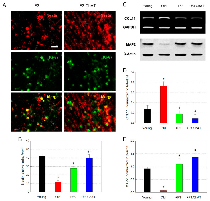
Major features of aging might be progressive decreases in cognitive function and physical activity, in addition to withered appearance. Previously, we reported that the intracerebroventricular injection of human neural stem cells (NSCs named F3) encoded the choline acetyltransferase gene (F3.ChAT). The cells secreted acetylcholine and growth factors (GFs) and neurotrophic factors (NFs), thereby improving learning and memory function as well as the physical activity of aged animals. In this study, F344 rats (10 months old) were intravenously transplanted with F3 or F3.ChAT NSCs (1 × 10⁶ cells) once a month to the 21st month of age. Their physical activity and cognitive function were investigated, and brain acetylcholine (ACh) and cholinergic and dopaminergic system markers were analyzed. Neuroprotective and neuroregenerative activities of stem cells were also confirmed by analyzing oxidative damages, neuronal skeletal protein, angiogenesis, brain and muscle weights, and proliferating host stem cells. Stem cells markedly improved both cognitive and physical functions, in parallel with the elevation in ACh levels in cerebrospinal fluid and muscles, in which F3.ChAT cells were more effective than F3 parental cells. Stem cell transplantation downregulated CCL11 and recovered GFs and NFs in the brain, leading to restoration of microtubule-associated protein 2 as well as functional markers of cholinergic and dopaminergic systems, along with neovascularization. Stem cells also restored muscular GFs and NFs, resulting in increased angiogenesis and muscle mass. In addition, stem cells enhanced antioxidative capacity, attenuating oxidative damage to the brain and muscles. The results indicate that NSCs encoding ChAT improve cognitive function and physical activity of aging animals by protecting and recovering functions of multiple organs, including cholinergic and dopaminergic systems, as well as muscles from oxidative injuries through secretion of ACh and GFs/NFs, increased antioxidant elements, and enhanced blood flow.
Keywords: acetylcholine; aging; choline acetyltransferase; cognitive function; growth factor; human neural stem cell; neurotrophic factor; physical activity.
Conflict of interest statement
Authors Tae Myoung Kim, Ehn-Kyoung Choi, and Yun-Bae Kim were employed by the company Designed Cells Co., Ltd. The remaining authors declare that the research was conducted in the absence of any commercial or financial relationships that could be constructed as a potential conflict of interest. The authors declare that this study received funding from National Research Foundation of Korea and Korea Health Industry Development Institute. The funders were not involved in the study design, collection, analysis, interpretation of data, the writing of this article or the decision to submit it for publication.
Figures


Similar articles
-
Improvement of cognitive function and physical activity of aging mice by human neural stem cells over-expressing choline acetyltransferase.Neurobiol Aging. 2013 Nov;34(11):2639-46. doi: 10.1016/j.neurobiolaging.2013.04.026. Epub 2013 May 31. Neurobiol Aging. 2013. PMID: 23731954
-
Human Neural Stem Cells Encoding ChAT Gene Restore Cognitive Function via Acetylcholine Synthesis, Aβ Elimination, and Neuroregeneration in APPswe/PS1dE9 Mice.Int J Mol Sci. 2020 May 31;21(11):3958. doi: 10.3390/ijms21113958. Int J Mol Sci. 2020. PMID: 32486466 Free PMC article.
-
Neuroprotective effects of human neural stem cells over-expressing choline acetyltransferase in a middle cerebral artery occlusion model.J Chem Neuroanat. 2020 Jan;103:101730. doi: 10.1016/j.jchemneu.2019.101730. Epub 2019 Dec 16. J Chem Neuroanat. 2020. PMID: 31837389
-
Recent preclinical evidence advancing cell therapy for Alzheimer's disease.Exp Neurol. 2012 Sep;237(1):142-6. doi: 10.1016/j.expneurol.2012.06.024. Epub 2012 Jun 27. Exp Neurol. 2012. PMID: 22766481 Free PMC article. Review.
-
Overexpression of choline acetyltransferase reconstitutes discrete acetylcholine release in some but not all synapse formation-defective neuroblastoma cells.J Physiol Paris. 1995;89(3):137-45. doi: 10.1016/0928-4257(96)80111-2. J Physiol Paris. 1995. PMID: 7581303 Review.
Cited by
-
Hippocampal Viral-Mediated Urokinase Plasminogen Activator (uPA) Overexpression Mitigates Stress-Induced Anxiety and Depression in Rats by Increasing Brain-Derived Neurotrophic Factor (BDNF) Levels.Biomolecules. 2024 Dec 15;14(12):1603. doi: 10.3390/biom14121603. Biomolecules. 2024. PMID: 39766310 Free PMC article.
-
The therapeutic potential of neuroglobin-overexpressing human neural stem cells in a photothrombosis model.Regen Ther. 2025 Aug 5;30:515-524. doi: 10.1016/j.reth.2025.07.013. eCollection 2025 Dec. Regen Ther. 2025. PMID: 40860426 Free PMC article.
References
-
- Vanguilder H.D., Bixler G.V., Sonntag W.E., Freeman W.M. Hippocampal expression of myelin-associated inhibitors is induced with age-related cognitive decline and correlates with deficits of spatial learning and memory. J. Neurochem. 2012;121:77–98. doi: 10.1111/j.1471-4159.2012.07671.x. - DOI - PMC - PubMed
Publication types
MeSH terms
Substances
Grants and funding
LinkOut - more resources
Full Text Sources
Miscellaneous
