Review
. 2023 Nov 29;12(23):2730.
doi: 10.3390/cells12232730.

Modulating Growth Factor Receptor Signaling to Promote Corneal Epithelial Homeostasis

Affiliations
Review

Modulating Growth Factor Receptor Signaling to Promote Corneal Epithelial Homeostasis

Kate E Tarvestad-Laise et al. Cells. .

Abstract

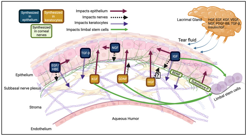
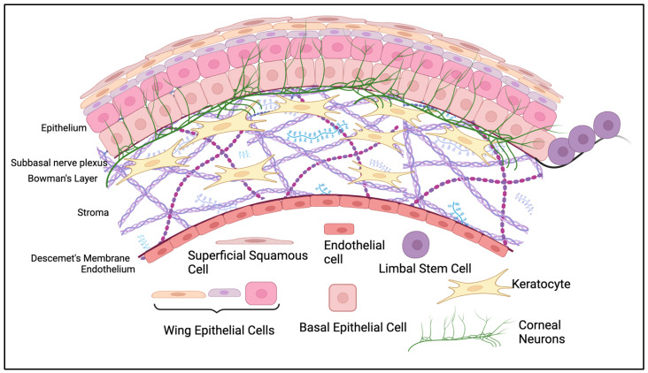
The corneal epithelium is the first anatomical barrier between the environment and the cornea; it is critical for proper light refraction onto the retina and prevents pathogens (e.g., bacteria, viruses) from entering the immune-privileged eye. Trauma to the highly innervated corneal epithelium is extremely painful and if not resolved quickly or properly, can lead to infection and ultimately blindness. The healthy eye produces its own growth factors and is continuously bathed in tear fluid that contains these proteins and other nutrients to maintain the rapid turnover and homeostasis of the ocular surface. In this article, we review the roles of growth factors in corneal epithelial homeostasis and regeneration and some of the limitations to their use therapeutically.

Keywords: c-Met; cornea; corneal epithelium; corneal nerves; growth factor; growth factor receptor; wound healing.


Conflict of interest statement

The authors declare no conflict of interest.

Figures

Figure 2
Figure 2
Growth factor expression in the corneal epithelium. A sample of some of the main mediators of corneal epithelial homeostasis. Many growth factors, like EGF, HB-EGF, NGF, and TGF-β are synthesized in the epithelium and act in an autocrine manner, where they bind to epithelial cells, or a paracrine manner, where they bind to nearby cells, like keratocytes or corneal nerves. Alternatively, proteins like HGF and KGF are expressed in keratocytes and diffuse anteriorly to the corneal nerves and epithelium. Some proteins synthesized in the cornea also act on the adjacent limbal stem cells to promote their proliferation. Corneal nerves release neurotrophic factors like substance P and brain derived neurotrophic factor (BDNF) that act in autocrine or paracrine manners [36]. Many of these growth factors can be synthesized in multiple layers of the cornea. Finally, growth factors can reach the corneal epithelium via the lacrimal gland and the tear fluid or through the aqueous humor (regulated by the endothelium). A more comprehensive list of growth factors is in Table 1. Created with BioRender.com (accessed on 17 November 2023).
Figure 3
Figure 3
Receptor desensitization. [Starting top left] Ligand induces the dimerization of two receptor monomers, causing conformational changes and leading to auto-transphosphorylation. Phosphorylated tyrosines serve as catalytic or docking sites (See Figure 4). Concomitantly, receptor dimers translocate to clathrin rich membrane domains which invaginate to form a clathrin-coated pit containing the ligand-receptor complex [203]. Dynamin pinches off the membrane and generates a clathrin-coated vesicle [204,205,206]. Clathrin proteins are shed and recycled. The resulting naked vesicle is delivered to and fuses with the early endosome. The early endosome sorts the cargo for its ultimate cellular fate. Receptors can be recycled back to the plasma membrane for additional rounds of activation. Alternatively, receptors can be retained in the early endosome which matures into a late endosome [200]. E3 ubiquitin ligases (i.e., c-Cbl, Cbl-b) bind and transfer ubiquitin to the receptor. Receptor ubiquitylation is a critical modification for endocytosis and allows the receptor to be recognized by the ESCRT complexes. Ubiquitylated receptors bind to ESCRT proteins and become sequestered into intralumenal vesicles (ILV) within the mature late endosome. The late endosome fuses with the lysosome and the cargo is transferred for degradation. Created with BioRender.com (accessed on 17 November 2023).
Figure 4
Figure 4
Example of the c-Met receptor’s activation pathways. Receptor activation begins with two HGF molecules binding to two c-Met monomers, promoting the dimerization and stabilization of the complex [228]. Dimerization of the transmembrane monomers allows for the kinase domains on each receptor to activate via auto-transphosphorylation. Once the catalytic domains are active (Y1234/Y1235), they mediate the phosphorylation of tyrosine residues in both the juxtamembrane (Y1003) and docking domains (Y1349/Y1356). The phosphorylated docking domain allows for scaffold (i.e., GAB1, Grb2) and effector proteins (i.e., STAT3, PI3K) to bind. Effector proteins can be phosphorylated directly by the receptor, or by binding onto scaffold proteins to be brought in range of the c-Met catalytic domain. Different outcomes are driven by which effector proteins are activated. A few pathways are clearer than others, namely the Ras/Raf/Mek/ERK1/2 pathway and its involvement in proliferation and cell motility [229]. It is also well established that PI3-K works through Akt to prevent apoptosis in corneal epithelial cells [230]. Other pathways, including the CRK-JNK for transformation [231,232] and STAT3 for invasion [233,234], have various results, so the outcome of signaling through these pathways may be determined by cell type. One gap in the research is what pathways are activated and how c-Met plays a role in nerve regeneration, preventing fibrosis, and angiogenesis. Following the phosphorylation of Y1003 in the juxtamembrane domain, E3 ubiquitin ligases like c-Cbl or Cbl-b can bind and transfer ubiquitin molecules to the receptor, ultimately ending with receptor degradation (see Figure 4). Created with BioRender.com (accessed on 17 November 2023).
Figure 1
Figure 1
Anatomical structure of the corneal epithelium. The most anterior epithelial layer is made of 5–7 layers of epithelial cells. Basal epithelial cells arise from limbal stem cells (LSCs) that move centripetally into the cornea from the limbus. As basal epithelial cells move through their life cycle, they become smaller, move anteriorly, and are eventually shed as superficial squamous cells. The epithelium lies above Bowman’s Layer and the subbasal plexus, which are anterior to the stroma. The stroma is the thickest layer and is mainly populated by keratocytes, which release extracellular matrix and collagen to maintain the transparency of the cornea. The stroma is separated from the most posterior layer of the cornea, the endothelium, by Descemet’s membrane. The endothelium is made up of a single layer of endothelial cells that tightly regulate fluid dynamics from the aqueous humor. Created with BioRender.com (accessed on 17 November 2023).

Similar articles

Cited by

References

    1. Scott P. Scott’s Anatomy of the Eye and Orbit. Ridgevue Publishing; Atascadero, CA, USA: 2019.
    1. Müller L.J., Marfurt C.F., Kruse F., Tervo T.M.T. Corneal nerves: Structure, contents and function. Exp. Eye Res. 2003;76:521–542. doi: 10.1016/S0014-4835(03)00050-2. - DOI - PubMed
    1. Koh S., Rao S.K., Srinivas S.P., Tong L., Young A.L. Evaluation of ocular surface and tear function—A review of current approaches for dry eye. Indian J. Ophthalmol. 2022;70:1883–1891. doi: 10.4103/ijo.IJO_1804_21. - DOI - PMC - PubMed
    1. Klenkler B., Sheardown H., Jones L. Growth factors in the tear film: Role in tissue maintenance, wound healing, and ocular pathology. Ocul. Surf. 2007;5:228–239. doi: 10.1016/S1542-0124(12)70613-4. - DOI - PubMed
    1. Bonanno J.A. Molecular mechanisms underlying the corneal endothelial pump. Exp. Eye Res. 2012;95:2–7. doi: 10.1016/j.exer.2011.06.004. - DOI - PMC - PubMed

Publication types

Substances

LinkOut - more resources