. 2023 Nov 29;12(23):2733.
doi: 10.3390/cells12232733.

Cytotoxic Effect Induced by Sicilian Oregano Essential Oil in Human Breast Cancer Cells

Affiliations

Cytotoxic Effect Induced by Sicilian Oregano Essential Oil in Human Breast Cancer Cells

Diana Di Liberto et al. Cells. .

Abstract

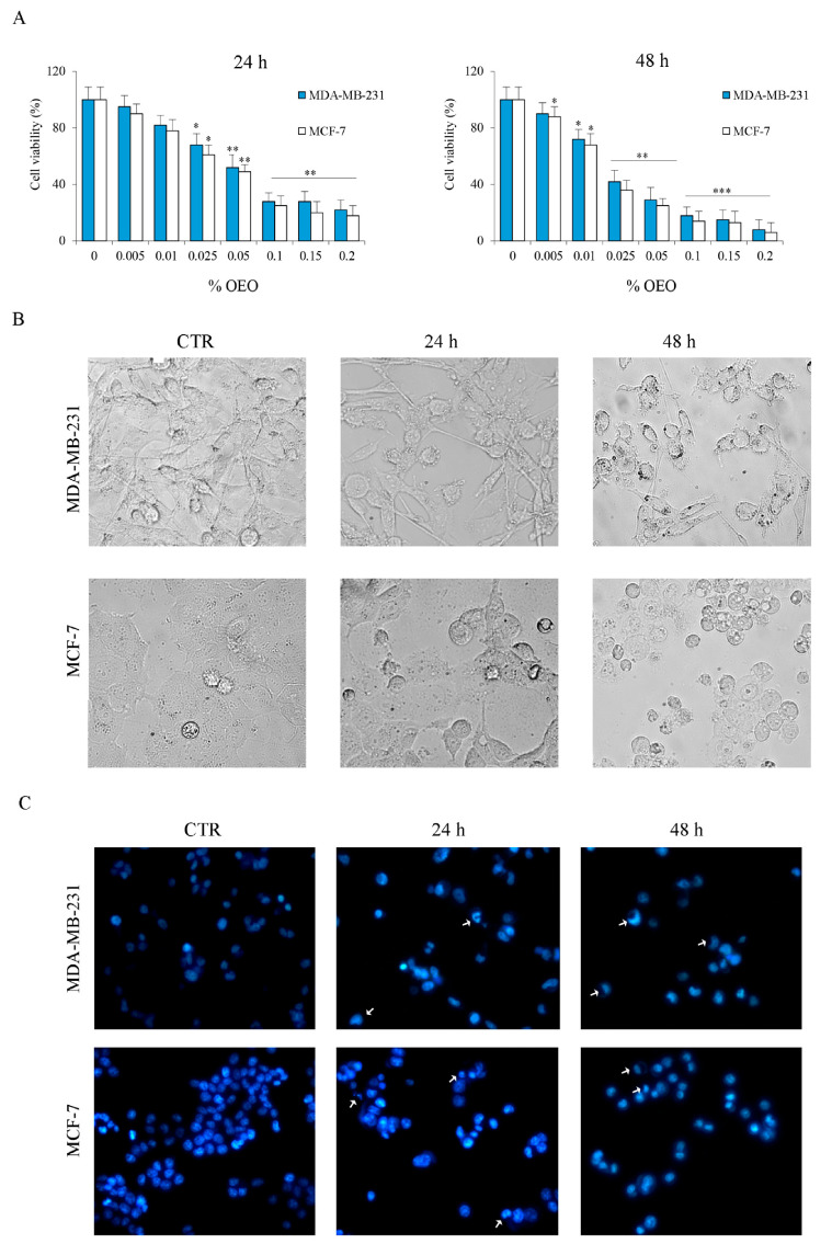
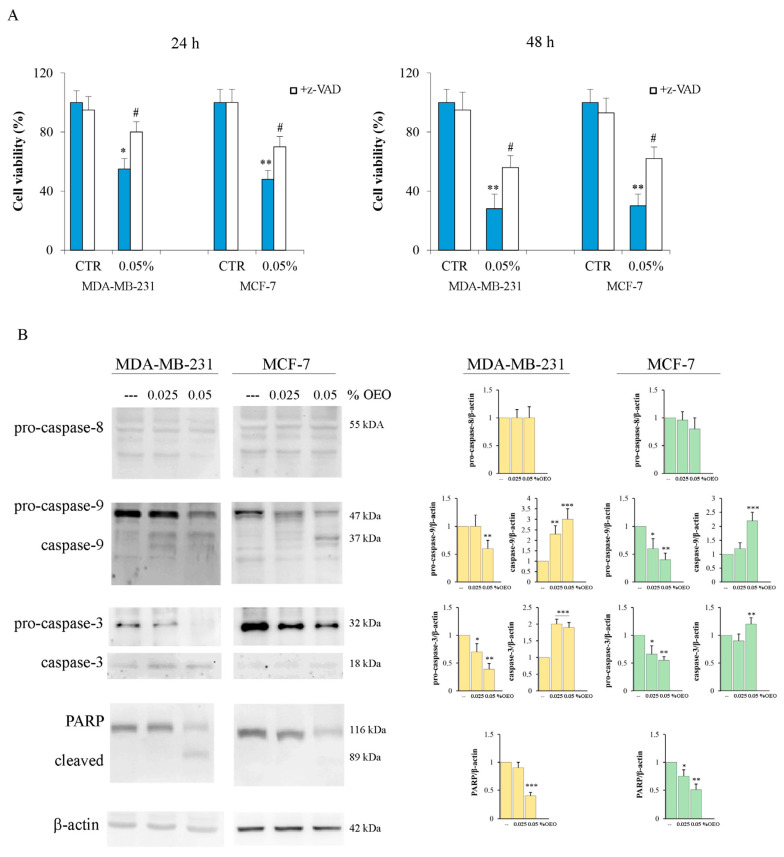
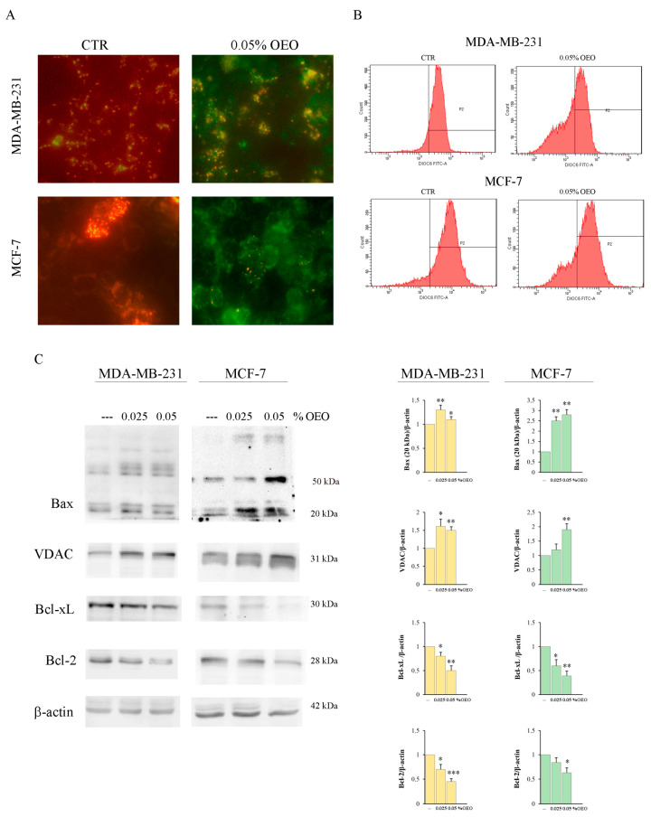
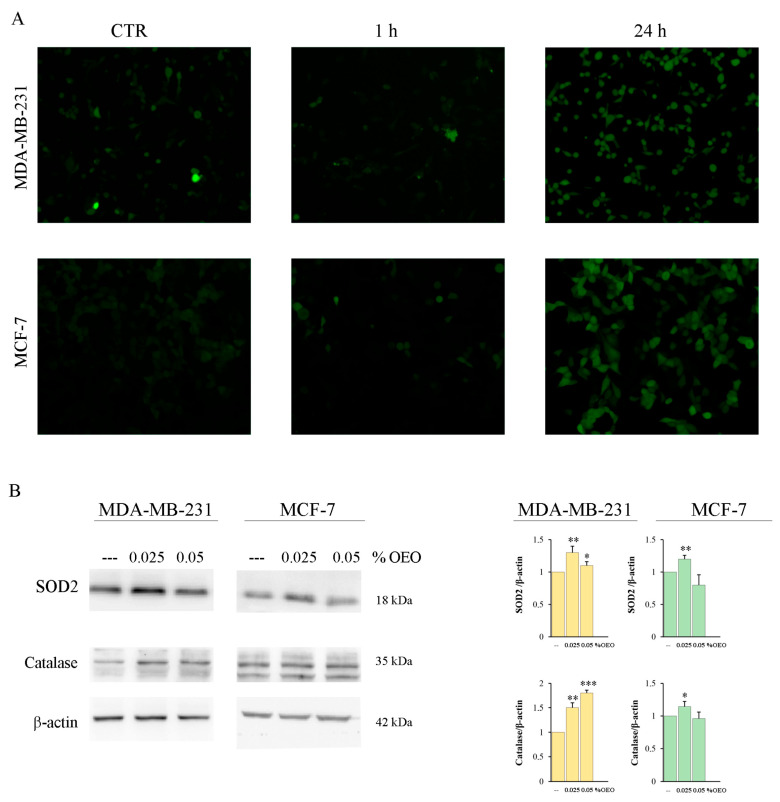
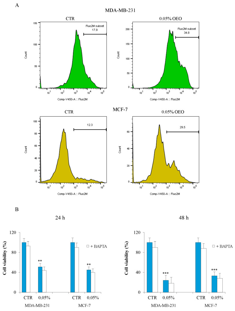
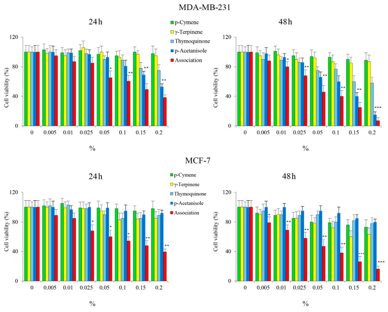
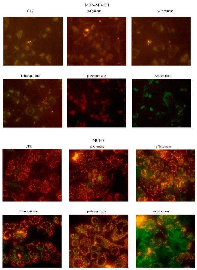
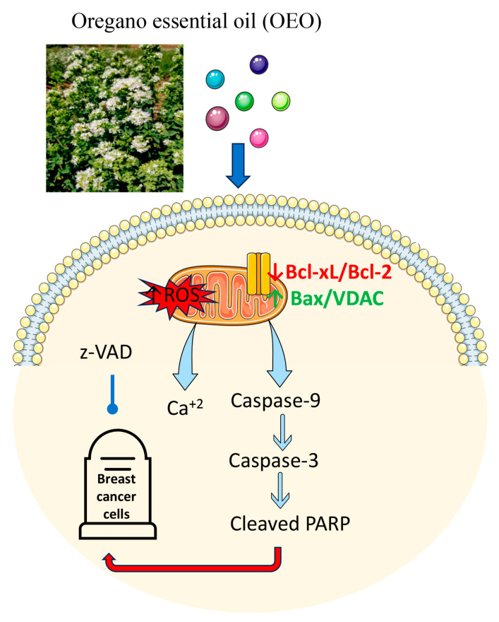
Origanum vulgare L. is an aromatic plant that exerts antibacterial, antioxidant, anti-inflammatory, and antitumor activities, mainly due to its essential oil (EO) content. In this study, we investigated the possible mechanism underlying the in vitro antitumor activity of EO extracted by hydrodistillation of dried flowers and leaves of Origanum vulgare L. grown in Sicily (Italy) in MDA-MB-231 and MCF-7 breast cancer cell lines. Gas chromatography-mass spectrometry analysis of Oregano essential oil (OEO) composition highlighted the presence of twenty-six major phytocompounds, such as p-cymene, γ-terpinene, and thymoquinone p-acetanisole. OEO possesses strong antioxidant capacity, as demonstrated by the DPPH test. Our studies provided evidence that OEO reduces the viability of both MCF-7 and MDA-MB-231 cells. The cytotoxic effect of OEO on breast cancer cells was partially counteracted by the addition of z-VAD-fmk, a general caspase inhibitor. Caspases and mitochondrial dysfunction appeared to be involved in the OEO-induced death mechanism. Western blotting analysis showed that OEO-induced activation of pro-caspases-9 and -3 and fragmentation of PARP decreased the levels of Bcl-2 and Bcl-xL while increasing those of Bax and VDAC. In addition, fluorescence microscopy and cytofluorimetric analysis showed that OEO induces a loss of mitochondrial membrane potential in both cell lines. Furthermore, we tested the effects of p-cymene, γ-terpinene, thymoquinone, and p-acetanisole, which are the main components of OEO. Our findings highlighted that the effect of OEO on MDA-MB-231 and MCF-7 cells appears to be mainly due to the combination of different constituents of OEO, providing evidence of the potential use of OEO for breast cancer treatment.

Keywords: GC–MS; antioxidant activity; breast cancer; essential oil; oregano.


Conflict of interest statement

The authors declare no conflict of interest.

Figures

Figure 1
Figure 1
The cytotoxic effect exerted by OEO on MDA-MB-231 and MCF-7 cells. (A) Effects of OEO on cell viability. Cells were treated with different percentages of OEO for 24 h and 48 h. Cell viability was assessed by MTT, as described in Section 2. (B) Morphologic changes in MDA-MB-231 and MCF-7 cells were observed under (B) light microscopy and (C) fluorescence microscopy after Hoechst33342 staining. Images were taken at 200X magnification. Arrows indicate OEO-induced changes in chromatin. Results are representative of three independent experiments. In (A), values are the means of three independent experiments ± S.D. * p < 0.05, ** p < 0.01, *** p < 0.001 vs. untreated control.
Figure 2
Figure 2
OEO-induced caspase activation. (A) The caspase inhibitor z-VAD-FMK reduced the OEO cytotoxic effect in BC cells. MDA-MB-231 and MCF-7 cells were treated with 0.05% OEO without or with 50 µM z-VAD-FMK for 24 h and 48 h. Cell viability was assessed by MTT, as described in Section 2. (B) The effect of OEO on pro-caspase activation. MDA-MB-231 and MCF-7 cells were treated for 24 h with 0.025 or 0.05% OEO and then evaluated for the level of pro-caspases 8, -9, and -3 and their cleavage forms by Western blotting analysis. The data show the densitometric analyses obtained using the Quantity One software. The protein content was normalized with respect to β-actin; the reported values, the mean of three independent experiments, are reported with respect to the untreated control, which was assigned a value of 1. In (A,B), values are the means of three independent experiments ± S.D. * p < 0.05, ** p < 0.01; *** p < 0.001 vs. untreated control; # p < 0.05 vs. treated cells.
Figure 3
Figure 3
The effects of OEO on mitochondrial membrane potential (ΔΨm) and levels of Bcl-2 family members and VDAC. (A,B) OEO-induced mitochondrial depolarization in MDA-MB-231 and MCF-7 cells. Cells were treated for 24 h with 0.05% OEO. (A) In the end, cells were incubated with JC-1 fluorochrome, and fluorescent cells were visualized with an OPTIKA microscope at 400X magnification, as indicated in Section 2. (B) Dissipation of ΔΨm was also evaluated by flow cytometry using the lipophilic dye DiOC6. (C) The effect of OEO on the BCl-2 family and VDAC. MDA-MB-231 and MCF-7 cells were treated for 24 h with 0.025 or 0.05% OEO, and then the levels of Bax, VDAC, Bcl-xL, and Bcl-2 were evaluated by Western blotting analysis. The data show the densitometric analyses obtained using the Quantity One software. The protein content was normalized with respect to β-actin; the reported values, the mean of three independent experiments, are reported with respect to the untreated control, which was assigned a value of 1. The results are representative of three independent experiments. In (C), the reported values are the means of three independent experiments ± S.D. * p < 0.05, ** p < 0.01; *** p < 0.001 vs. untreated control.
Figure 4
Figure 4
The effects of OEO on oxidative stress. (A) OEO induces ROS production. MDA-MB-231 and MCF-7 were treated for 1 h and 24 h with 0.05% OEO. In the end, cells were incubated with 1 µM H2-DCFDA, and fluorescent cells were visualized with an OPTIKA microscope at 200X magnification, as indicated in Section 2. (B) The effect of OEO on antioxidant enzymes. MDA-MB-231 and MCF-7 cells were treated for 24 h with 0.025 or 0.05% OEO, and then the levels of SOD2 and catalase were evaluated with Western blotting analysis. The data show the densitometric analyses obtained using the Quantity One software. The protein content was normalized with respect to β-actin; the reported values, the mean of three independent experiments, are reported with respect to the untreated control, which was assigned a value of 1. (A) The results are representative of three independent experiments. In (B), the reported values are the means of three independent experiments ± S.D. * p < 0.05, ** p < 0.01; *** p < 0.001 vs. untreated control.
Figure 5
Figure 5
OEO treatment increased the intracellular calcium level. (A) MDA-MB-231 and MCF-7 cells were treated with 0.05% OEO for 24 h and 48 h. Variation in the content of intracellular calcium was evaluated by flow cytometry using Fluo 2-AM fluorochrome as reported in Section 2. (B) Effects of OEO/BAPTA treatment on cell viability. Cells were treated with 0.05% OEO without or with 5 µM BAPTA for 24 h and 48 h. Cell viability was assessed by MTT, as described in Section 2. In (B), values are the means of three independent experiments ± S.D. ** p < 0.01; *** p < 0.001 vs. untreated control.
Figure 6
Figure 6
The cytotoxic effect exerted by some constituents of OEO on MDA-MB-231 and MCF-7 cells. Cells were treated with different percentages of p-cymene, γ-terpinene, thymoquinone, and p-acetanisole, employed alone or in association, for 24 h and 48 h. Cell viability was assessed by MTT, as described in Section 2. Results are representative of three independent experiments. Values are the means of three independent experiments ± S.D. * p < 0.05, ** p < 0.01, *** p < 0.001 vs. untreated control.
Figure 7
Figure 7
The effects of some constituents of OEO on mitochondrial membrane potential (ΔΨm). Cells were treated with p-cymene, γ-terpinene, thymoquinone, and p-acetanisole, employed alone or in association, for 24 h. In the end, cells were incubated with JC-1 fluorochrome, and fluorescent cells were visualized with an OPTIKA microscope at 400X magnification, as indicated in Section 2.
Figure 8
Figure 8
Schematic representation of the mechanism of action of OEO. Parts of the figure were drawn by using pictures from Servier Medical Art. Servier Medical Art by Servier is licensed under a Creative Commons Attribution 3.0 Unported License (https://creativecommons.org/licenses/by/3.0/ accessed on 19 September 2023).

Similar articles

Cited by

References

    1. Arabaci T., Çelenk S., Özcan T., Martin E., Yazici T., Açar M., Üzel D., Dirmenci T. Homoploid Hybrids of Origanum (Lamiaceae) in Turkey: Morphological and Molecular Evidence for a New Hybrid. Plant Biosyst. Int. J. Deal. All Asp. Plant Biol. 2021;155:470–482. doi: 10.1080/11263504.2020.1762777. - DOI
    1. Harborne M.S., Jeffrey B. Oregano. CRC Press; Boca Raton, FL, USA: 2002. The Taxonomy and Chemistry of Origanum.
    1. Lotti C., Ricciardi L., Rainaldi G., Ruta C., Tarraf W., De Mastro G. Morphological, Biochemical, and Molecular Analysis of Origanum vulgare L. Open Agric. J. 2019;13:116–124. doi: 10.2174/1874331501913010116. - DOI
    1. Lombrea A., Antal D., Ardelean F., Avram S., Pavel I.Z., Vlaia L., Mut A.-M., Diaconeasa Z., Dehelean C.A., Soica C., et al. A Recent Insight Regarding the Phytochemistry and Bioactivity of Origanum vulgare L. Essential Oil. Int. J. Mol. Sci. 2020;21:9653. doi: 10.3390/ijms21249653. - DOI - PMC - PubMed
    1. Bakkali F., Averbeck S., Averbeck D., Idaomar M. Biological Effects of Essential Oils—A Review. Food Chem. Toxicol. 2008;46:446–475. doi: 10.1016/j.fct.2007.09.106. - DOI - PubMed

Publication types