. 2023 Nov 30;12(23):2736.
doi: 10.3390/cells12232736.

Alteration of Immunoregulatory Patterns and Survival Advantage of Key Cell Types in Food Allergic Children

Affiliations

Alteration of Immunoregulatory Patterns and Survival Advantage of Key Cell Types in Food Allergic Children

Kamal Ivory et al. Cells. .

Abstract

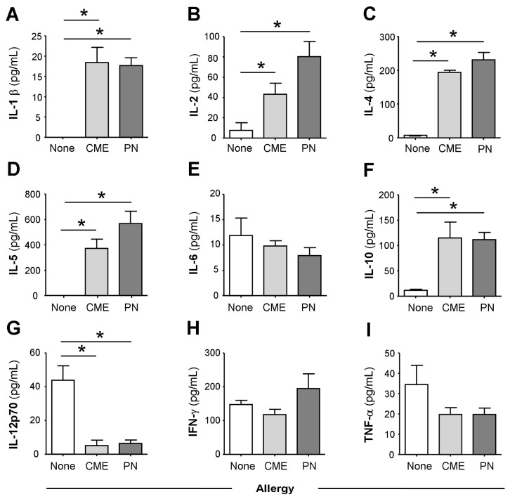
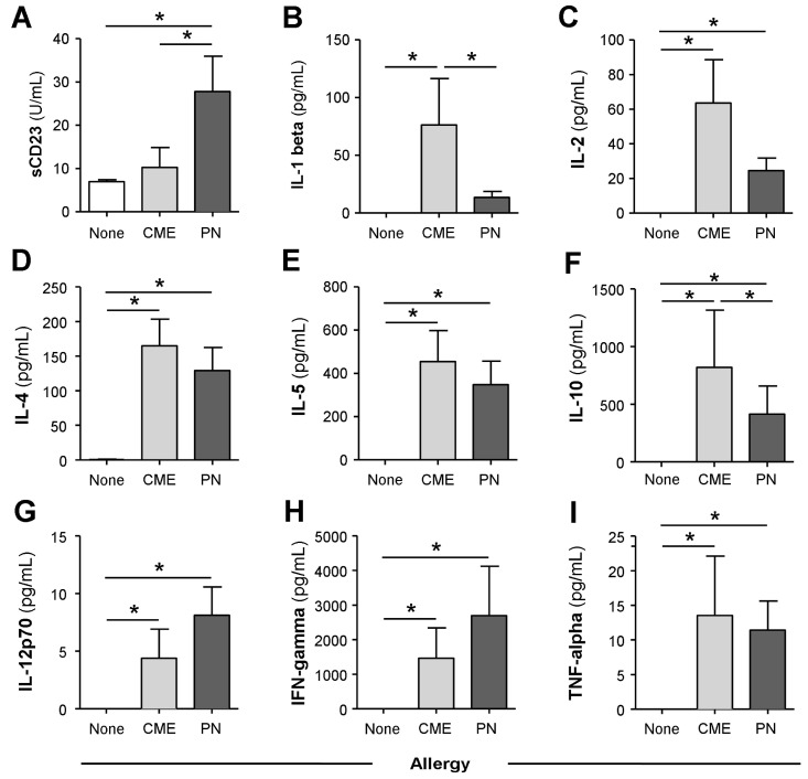
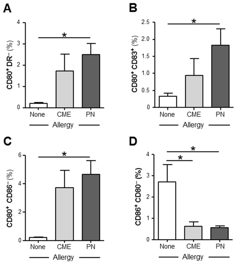
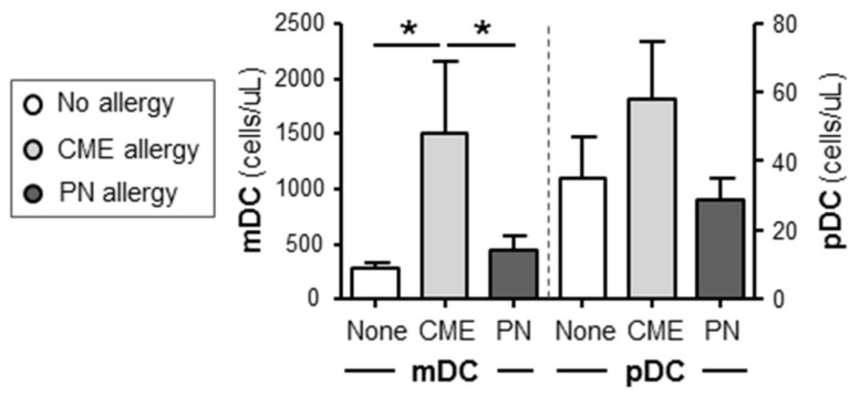
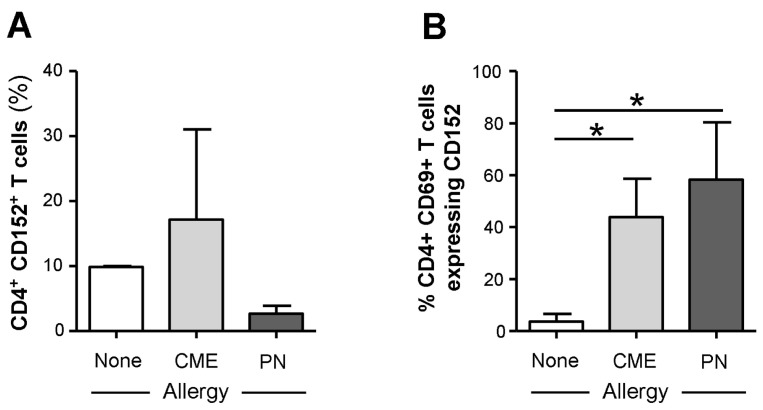
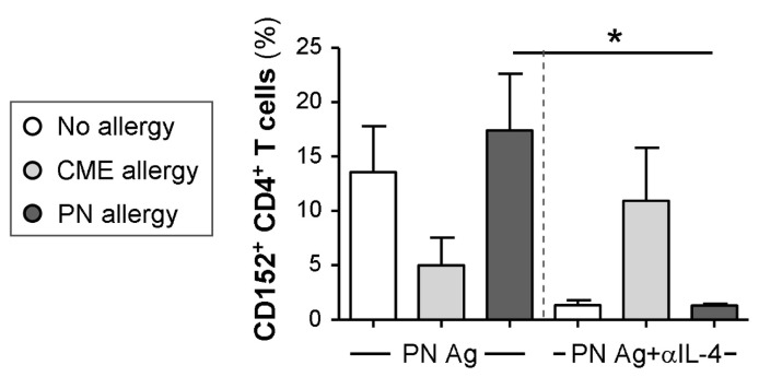
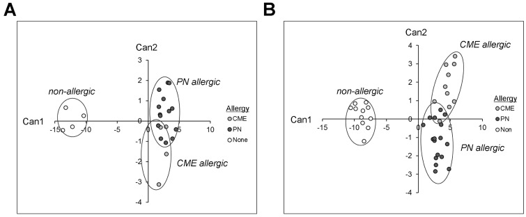
All allergic responses to food indicate the failure of immunological tolerance, but it is unclear why cow's milk and egg (CME) allergies resolve more readily than reactivity to peanuts (PN). We sought to identify differences between PN and CME allergies through constitutive immune status and responses to cognate and non-cognate food antigens. Children with confirmed allergy to CME (n = 6) and PN (n = 18) and non-allergic (NA) (n = 8) controls were studied. Constitutive secretion of cytokines was tested in plasma and unstimulated mononuclear cell (PBMNC) cultures. Blood dendritic cell (DC) subsets were analyzed alongside changes in phenotypes and soluble molecules in allergen-stimulated MNC cultures with or without cytokine neutralization. We observed that in allergic children, constitutively high plasma levels IL-1β, IL-2, IL-4, IL-5 and IL-10 but less IL-12p70 than in non-allergic children was accompanied by the spontaneous secretion of sCD23, IL-1β, IL-2, IL-4, IL-5, IL-10, IL-12p70, IFN-γ and TNF-α in MNC cultures. Furthermore, blood DC subset counts differed in food allergy. Antigen-presenting cell phenotypic abnormalities were accompanied by higher B and T cell percentages with more Bcl-2 within CD69+ subsets. Cells were generally refractory to antigenic stimulation in vitro, but IL-4 neutralization led to CD152 downregulation by CD4+ T cells from PN allergic children responding to PN allergens. Canonical discriminant analyses segregated non-allergic and allergic children by their cytokine secretion patterns, revealing differences and areas of overlap between PN and CME allergies. Despite an absence of recent allergen exposure, indication of in vivo activation, in vitro responses independent of challenging antigen and the presence of unusual costimulatory molecules suggest dysregulated immunity in food allergy. Most importantly, higher Bcl-2 content within key effector cells implies survival advantage with the potential to mount abnormal responses that may give rise to the manifestations of allergy. Here, we put forward the hypothesis that the lack of apoptosis of key immune cell types might be central to the development of food allergic reactions.

Keywords: apoptosis; cytokines; food allergy; immunoregulation; pediatric allergy.


Conflict of interest statement

The authors declare no conflict of interest.

Figures

Figure 1
Figure 1
Constitutive plasma cytokines. (A) IL-1β, (B) IL-2, (C) IL-4, (D) IL-5, (E) IL-6, (F) IL-10, (G) IL-12p70, (H) IFN-γ and (I) TNF-α were evaluated using multiplex bead technology in the plasma of non-allergic children and those with CME or PN allergies. Data were acquired via flow cytometry and are shown as means ± s.e.m; * p < 0.05, analyzed using ANOVA with adjustment for multiple comparisons using Tukey.
Figure 2
Figure 2
Spontaneous in vitro sCD23 and cytokine secretion. MNCs from non-allergic children or those with CME or PN allergies were cultured for six days without allergenic stimulation. Culture supernatants were collected and their levels of (A) sCD23 were detected using ELISA, while cytokines (B) IL-1β, (C) IL-2, (D) IL-4, (E) IL-5, (F) IL-10, (G) IL-12p70, (H) IFN-γ and (I) TNF-α were measured using multiplex bead technology and flow cytometry. Data are shown as means ± s.e.m; * p < 0.05, analyzed using ANOVA with adjustment for multiple comparisons using Tukey.
Figure 3
Figure 3
Expression of costimulatory molecules. MNCs from non-allergic children and those with CME or PN allergies were cultured without allergenic stimulation. On day 6, cells were harvested, washed and stained with CD80-FITC, CD83-PE, CD86-APC and HLA-DR-ECD. Data for the percentages of (A) CD80+HLA DR, (B) CD80+ CD83+, (C) CD80+ CD86 and (D) CD86+ CD80 were acquired via flow cytometry and are shown as means ± s.e.m; * p < 0.05, analyzed using ANOVA with adjustment for multiple comparisons using Tukey.
Figure 4
Figure 4
Dendritic cell subset enumeration. Dendritic cells were quantified in heparinized whole blood from non-allergic children and those with CME or PN allergies. Data were acquired via flow cytometry and are shown as means ± s.e.m; * p < 0.05, analyzed using ANOVA with adjustment for multiple comparisons using Tukey.
Figure 5
Figure 5
Percentages of B cells, T cells, APCs and their Bcl-2 content. MNCs from non-allergic children and those with CME or PN allergies were first stained with each of two panels for cell surface antigens, both containing CD69-APC, CD80-FITC, CD83-FITC and CD86-FITC except that Panel 1 had CD3-ECD and Panel 2 had CD19-ECD as lineage marker. This was followed by intracellular staining with Bcl-2-PE. Percentages of (A) B cells, (B) T cells and (C) APC are shown alongside MFI values for Bcl-2 content within CD69+ (D) B cells, (E) T cells and (F) APC. All data were acquired via flow cytometry and are seen as means ± s.e.m; * p < 0.05, analyzed using ANOVA with adjustment for multiple comparisons using Tukey.
Figure 6
Figure 6
Cell surface expression of CD152 within CD69+ CD4+ T cells. MNCs from non-allergic children and those with CME or PN allergies were stained with CD3-FITC, CD152-PE, CD4-ECD and CD69-APC. Cells were gated using scatter profiles and CD3 expression to identify T cells. Data for the percentages of CD152+ cells within (A) CD4+ or (B) CD4+ CD69+ were acquired via flow cytometry and are shown as means ± s.e.m; * p < 0.05, analyzed using ANOVA with adjustment for multiple comparisons using Tukey.
Figure 7
Figure 7
Cytokine-mediated regulation of CD152 expression. MNCs from non-allergic children and those with CME or PN allergies were cultured in the presence or absence of PN antigens (Ag) and with or without an IL-4 neutralizing antibody. On day 6, cells were harvested, washed and stained with CD3-FITC, CD152-PE and CD4-ECD. Data for the percentages of positive cells were acquired via flow cytometry and are shown as means ± s.e.m; * p < 0.05, analyzed using ANOVA with adjustment for multiple comparisons using Tukey.
Figure 8
Figure 8
Canonical coefficients of non-allergic individuals and individuals allergic to CME or PN. Linear discriminant functions were based on cytokines secreted (A) in vivo and therefore constitutively present in plasma or (B) released following 6-day MNC culture in vitro.

References

    1. Ling Z., Li Z., Liu X., Cheng Y., Luo Y., Tong X., Yuan L., Wang Y., Sun J., Li L., et al. Altered Fecal Microbiota Composition Associated with Food Allergy in Infants. Appl. Environ. Microbiol. 2014;80:2546–2554. doi: 10.1128/AEM.00003-14. - DOI - PMC - PubMed
    1. Tomicić S., Fälth-Magnusson K., Böttcher M.F. Dysregulated Th1 and Th2 responses in food-allergic children—Does elimination diet contribute to the dysregulation? Pediatr. Allergy Immunol. 2010;4:649–655. doi: 10.1111/j.1399-3038.2009.00937.x. - DOI - PubMed
    1. Chatila T. Regulatory T cells: Key Players in Tolerance and Autoimmunity. Endocrinol. Metab. Clin. N. Am. 2009;38:265–272. doi: 10.1016/j.ecl.2009.01.002. - DOI - PMC - PubMed
    1. Kwon H.K., Lee C.G., So J.S., Chae C.S., Hwang J.S., Sahoo A., Nam J.H., Rhee J.H., Hwang K.C., Im S.H. Generation of regulatory dendritic cells and CD4+Foxp3+ T cells by probiotics administration suppresses immune disorders. Proc. Natl. Acad. Sci. USA. 2010;107:2159–2164. doi: 10.1073/pnas.0904055107. - DOI - PMC - PubMed
    1. Ruiter B., Shreffler W.G. The role of dendritic cells in food allergy. J. Allergy Clin. Immunol. 2012;129:921–928. doi: 10.1016/j.jaci.2012.01.080. - DOI - PubMed

Publication types