

A New Era of Integration between Multiomics and Spatio-Temporal Analysis for the Translation of EMT towards Clinical Applications in Cancer
- PMID: 38067168
- PMCID: PMC10706093
- DOI: 10.3390/cells12232740
A New Era of Integration between Multiomics and Spatio-Temporal Analysis for the Translation of EMT towards Clinical Applications in Cancer
Abstract
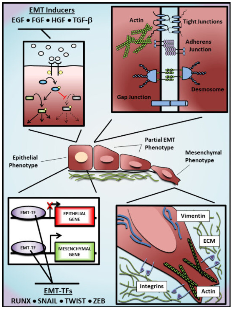
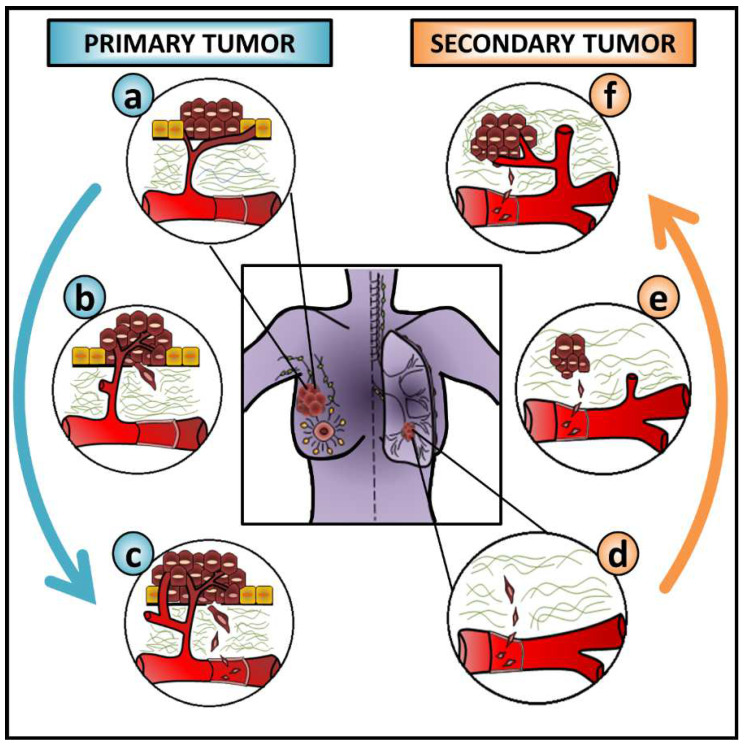
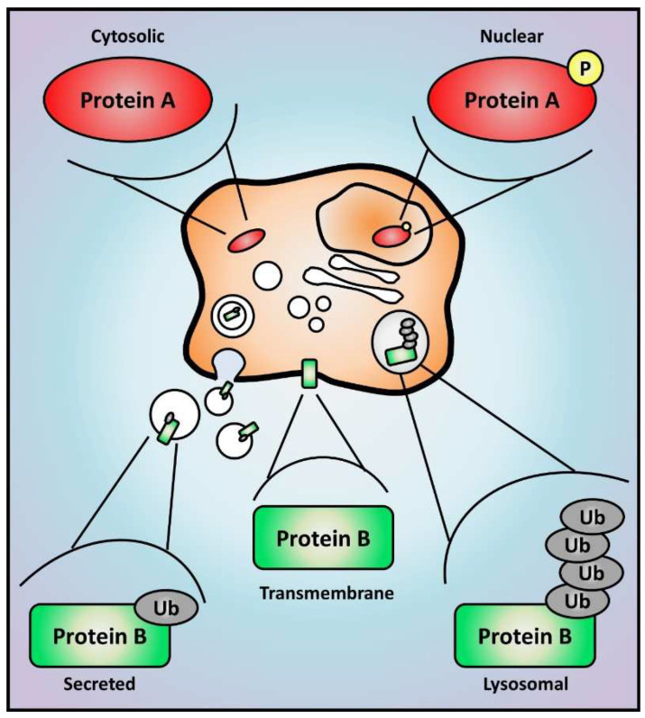
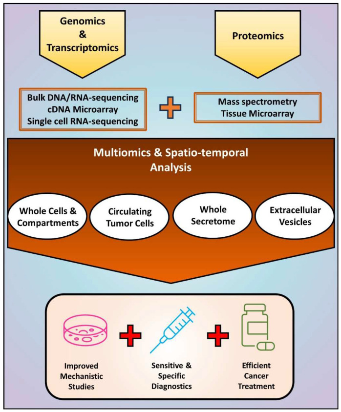
Epithelial-mesenchymal transition (EMT) is crucial to metastasis by increasing cancer cell migration and invasion. At the cellular level, EMT-related morphological and functional changes are well established. At the molecular level, critical signaling pathways able to drive EMT have been described. Yet, the translation of EMT into efficient diagnostic methods and anti-metastatic therapies is still missing. This highlights a gap in our understanding of the precise mechanisms governing EMT. Here, we discuss evidence suggesting that overcoming this limitation requires the integration of multiple omics, a hitherto neglected strategy in the EMT field. More specifically, this work summarizes results that were independently obtained through epigenomics/transcriptomics while comprehensively reviewing the achievements of proteomics in cancer research. Additionally, we prospect gains to be obtained by applying spatio-temporal multiomics in the investigation of EMT-driven metastasis. Along with the development of more sensitive technologies, the integration of currently available omics, and a look at dynamic alterations that regulate EMT at the subcellular level will lead to a deeper understanding of this process. Further, considering the significance of EMT to cancer progression, this integrative strategy may enable the development of new and improved biomarkers and therapeutics capable of increasing the survival and quality of life of cancer patients.
Keywords: EMT; cancer metastasis; genomics; multiomics; proteomics; secretome; transcriptomic.
Conflict of interest statement
The authors declare no conflict of interest. A.F.T., S.W., R.L. and H.-J.Z. are members of the research team at the Huagene Institute. Huagene Institute had no role in the design of the study; in the collection, analyses, or interpretation of data; in the writing of the manuscript, or in the decision to publish the results.
Figures
Similar articles
-
Proteomics analysis reveals the role of ubiquitin specific protease (USP47) in Epithelial to Mesenchymal Transition (EMT) induced by TGFβ2 in breast cells.J Proteomics. 2020 May 15;219:103734. doi: 10.1016/j.jprot.2020.103734. Epub 2020 Mar 19. J Proteomics. 2020. PMID: 32201364
-
EMTome: a resource for pan-cancer analysis of epithelial-mesenchymal transition genes and signatures.Br J Cancer. 2021 Jan;124(1):259-269. doi: 10.1038/s41416-020-01178-9. Epub 2020 Dec 10. Br J Cancer. 2021. PMID: 33299129 Free PMC article.
-
Integrated multi-omics analyses and functional validation reveal TTK as a novel EMT activator for endometrial cancer.J Transl Med. 2023 Feb 25;21(1):151. doi: 10.1186/s12967-023-03998-8. J Transl Med. 2023. PMID: 36829176 Free PMC article.
-
Multiomics technologies: role in disease biomarker discoveries and therapeutics.Brief Funct Genomics. 2023 Apr 13;22(2):76-96. doi: 10.1093/bfgp/elac017. Brief Funct Genomics. 2023. PMID: 35809340 Review.
-
Tumor microenvironment and noncoding RNAs as co-drivers of epithelial-mesenchymal transition and cancer metastasis.Dev Dyn. 2018 Mar;247(3):405-431. doi: 10.1002/dvdy.24548. Epub 2017 Sep 18. Dev Dyn. 2018. PMID: 28691356 Review.
Cited by
-
Factors Determining Epithelial-Mesenchymal Transition in Cancer Progression.Int J Mol Sci. 2024 Aug 17;25(16):8972. doi: 10.3390/ijms25168972. Int J Mol Sci. 2024. PMID: 39201656 Free PMC article. Review.
-
Extracellular Vesicles Secreted by Cancer-Associated Fibroblasts Drive Non-Invasive Cancer Cell Progression to Metastasis via TGF-β Signalling Hyperactivation.J Extracell Vesicles. 2025 Mar;14(3):e70055. doi: 10.1002/jev2.70055. J Extracell Vesicles. 2025. PMID: 40091448 Free PMC article.
-
A Novel Method for the Early Detection of Single Circulating, Metastatic and Self-Seeding Cancer Cells in Orthotopic Breast Cancer Mouse Models.Cells. 2024 Jul 9;13(14):1166. doi: 10.3390/cells13141166. Cells. 2024. PMID: 39056749 Free PMC article.
-
Enhancing detection and monitoring of circulating tumor cells: Integrative approaches in liquid biopsy advances.J Liq Biopsy. 2025 Apr 29;8:100297. doi: 10.1016/j.jlb.2025.100297. eCollection 2025 Jun. J Liq Biopsy. 2025. PMID: 40453103 Free PMC article. Review.
References
Publication types
MeSH terms
LinkOut - more resources
Full Text Sources
Medical
Miscellaneous
