. 2023 Dec 3;12(23):2759.
doi: 10.3390/cells12232759.

Role of Phosphodiesterase 1 in the Regulation of Real-Time cGMP Levels and Contractility in Adult Mouse Cardiomyocytes

Affiliations

Role of Phosphodiesterase 1 in the Regulation of Real-Time cGMP Levels and Contractility in Adult Mouse Cardiomyocytes

Nadja I Bork et al. Cells. .

Abstract

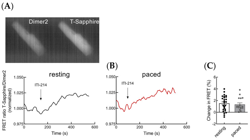
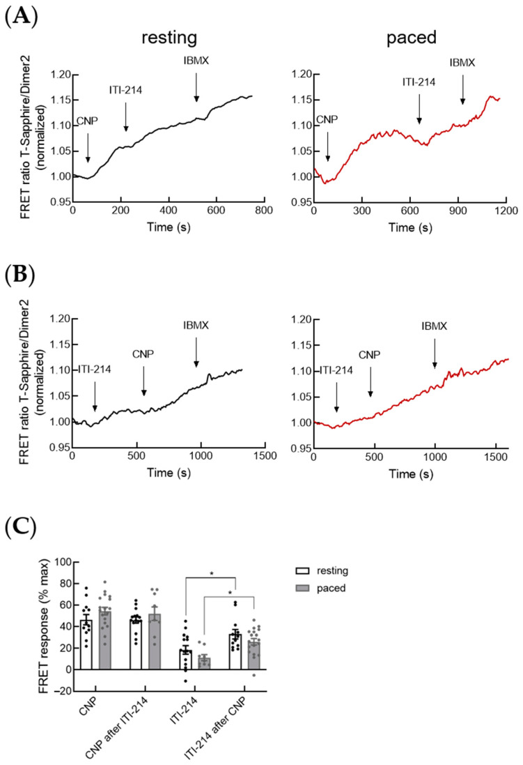
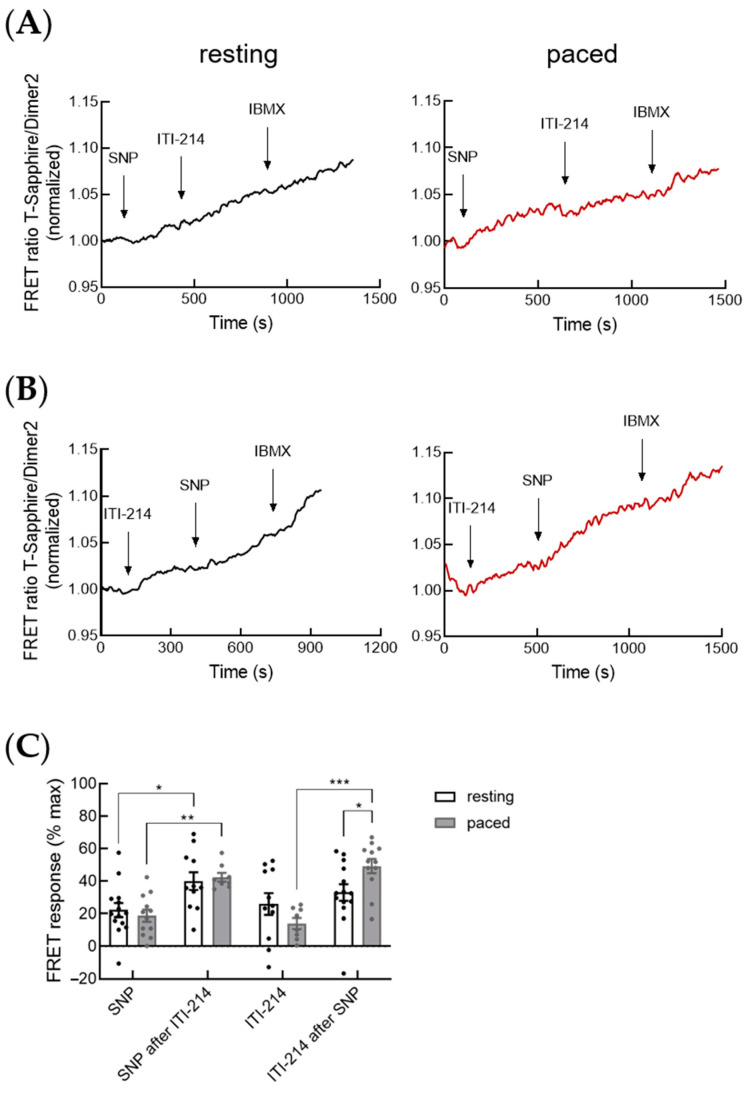
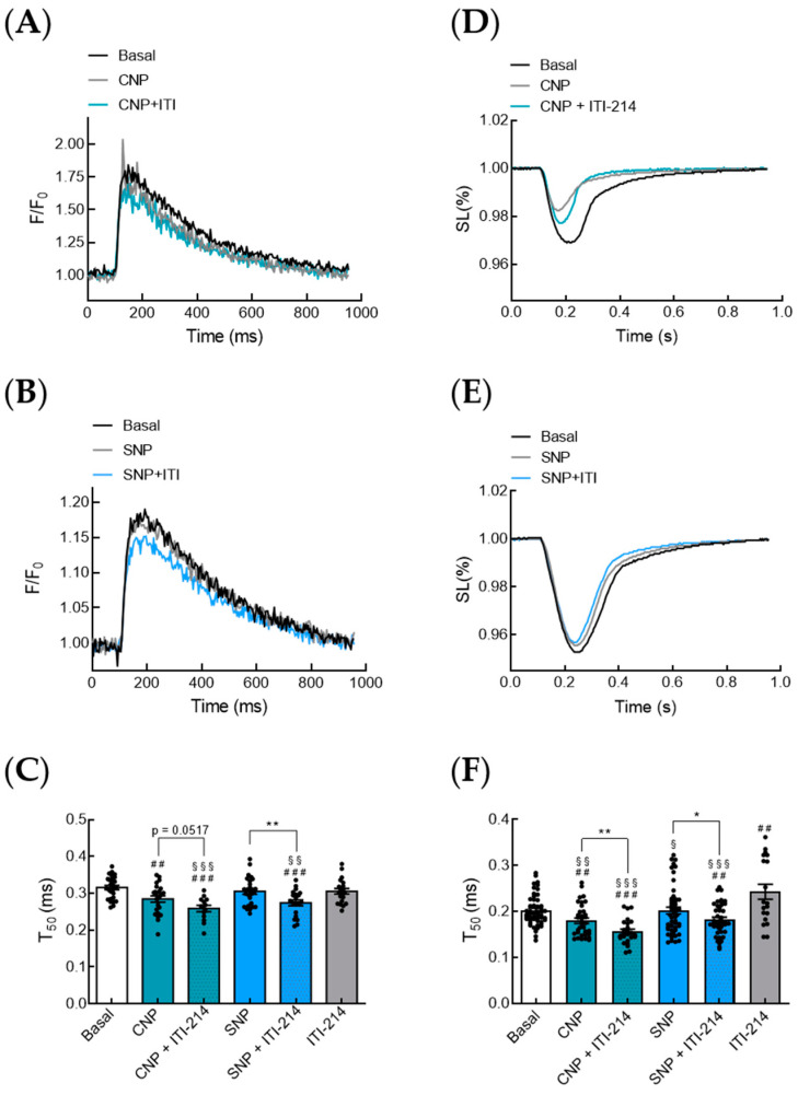
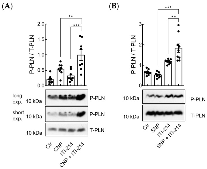
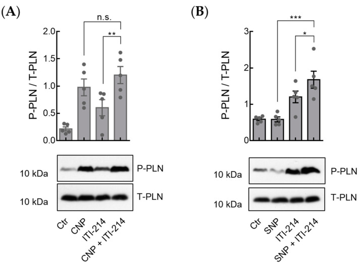
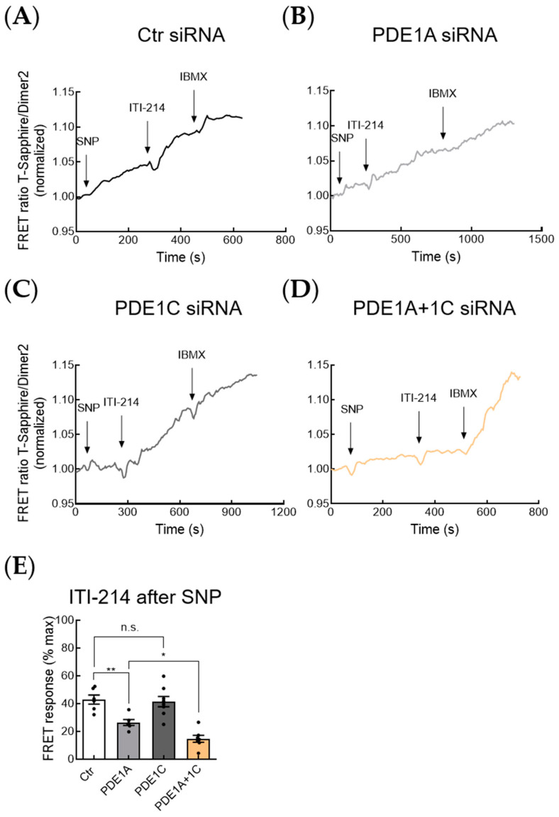
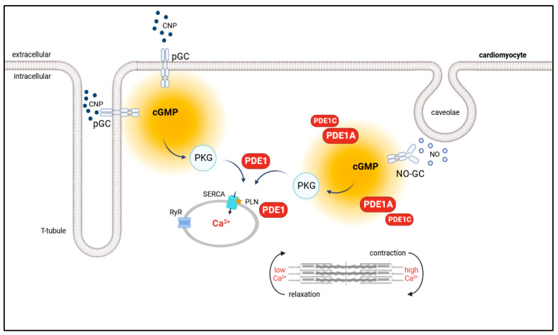
In mouse cardiomyocytes, the expression of two subfamilies of the calcium/calmodulin-regulated cyclic nucleotide phosphodiesterase 1 (PDE1)-PDE1A and PDE1C-has been reported. PDE1C was found to be the major subfamily in the human heart. It is a dual substrate PDE and can hydrolyze both 3',5'-cyclic adenosine monophosphate (cAMP) and 3',5'-cyclic guanosine monophosphate (cGMP). Previously, it has been reported that the PDE1 inhibitor ITI-214 shows positive inotropic effects in heart failure patients which were largely attributed to the cAMP-dependent protein kinase (PKA) signaling. However, the role of PDE1 in the regulation of cardiac cGMP has not been directly addressed. Here, we studied the effect of PDE1 inhibition on cGMP levels in adult mouse ventricular cardiomyocytes using a highly sensitive fluorescent biosensor based on Förster resonance energy transfer (FRET). Live-cell imaging in paced and resting cardiomyocytes showed an increase in cGMP after PDE1 inhibition with ITI-214. Furthermore, PDE1 inhibition and PDE1A knockdown amplified the cGMP-FRET responses to the nitric oxide (NO)-donor sodium nitroprusside (SNP) but not to the C-type natriuretic peptide (CNP), indicating a specific role of PDE1 in the regulation of the NO-sensitive guanylyl cyclase (NO-GC)-regulated cGMP microdomain. ITI-214, in combination with CNP or SNP, showed a positive lusitropic effect, improving the relaxation of isolated myocytes. Immunoblot analysis revealed increased phospholamban (PLN) phosphorylation at Ser-16 in cells treated with a combination of SNP and PDE1 inhibitor but not with SNP alone. Our findings reveal a previously unreported role of PDE1 in the regulation of the NO-GC/cGMP microdomain and mouse ventricular myocyte contractility. Since PDE1 serves as a cGMP degrading PDE in cardiomyocytes and has the highest hydrolytic activities, it can be expected that PDE1 inhibition might be beneficial in combination with cGMP-elevating drugs for the treatment of cardiac diseases.

Keywords: FRET; PDE1; cGMP; cardiomyocyte.


Conflict of interest statement

The authors declare no conflict of interest.

Figures

Figure 1
Figure 1
PDE1 inhibition increases cGMP-FRET in murine ventricular myocytes. (A,B) Representative raw fluorescence cell images in both fluorophore channels of the red-cGES-DE5 sensor and cGMP-FRET responses to PDE1 inhibition with ITI-214 (1 µM) alone. Representative traces showing the effect in resting (A) and paced (B) myocytes. (C) Quantification of cGMP-FRET responses to basal PDE1 inhibition shown in (A,B). Number of measured cardiomyocytes/mice were as follows: resting, 26/9; paced, 17/7.
Figure 2
Figure 2
PDE1 is not directly involved in the regulation of the cGMP pool generated by particulate guanylyl cyclase. (A) Representative FRET traces for the cGMP-FRET response in resting and paced myocytes to PDE1 inhibition with ITI-214 (1 µM) after stimulation with natriuretic peptide CNP (30 nM) followed by the pan-PDE inhibitor IBMX (100 µM). (B) Representative FRET traces for the cGMP-FRET response in resting and paced myocytes treated with ITI-214 (1 µM) followed by CNP (30 nM) and IBMX (100 µM). (C) Quantification of the FRET responses shown in (A,B). Number of measured cardiomyocytes/mice were as follows: CNP resting and ITI-214 after CNP resting = 12/6; CNP paced and ITI-214 after CNP paced = 18/4; ITI-214 resting and CNP after ITI-214 resting = 15/5; ITI-214 paced and CNP after ITI-214 paced = 9/3. Data in (C) were analyzed using nested t-test, * p < 0.05.
Figure 3
Figure 3
PDE1 is involved in the regulation of the cGMP pool generated by NO-GC. (A) Representative FRET traces for the cGMP-FRET response in resting and paced myocytes to PDE1 inhibition with ITI-214 (1 µM) after stimulation with NO-donor sodium nitroprusside (SNP, 50 µM), followed by the pan-PDE inhibitor IBMX (100 µM). (B) Representative FRET traces for cGMP-FRET response in resting and paced myocytes treated with ITI-214 (1 µM) followed by SNP (50 µM) and IBMX (100 µM). (C) Quantification of FRET responses shown in (A,B). Number of measured cardiomyocytes/mice were as follows: SNP resting and ITI-214 after SNP resting = 14/4; SNP paced and ITI-214 after SNP paced = 13/5; ITI-214 resting and SNP after ITI-214 resting = 11/4; ITI-214 paced and SNP after ITI-214 paced = 8/4. Data in (C) were analyzed via a nested t-test, * p < 0.05, ** p < 0.01, *** p < 0.001.
Figure 4
Figure 4
ITI-214 in combination with CNP or SNP shows positive lusitropic effects in isolated myocytes. (A) Representative Ca2+ transients from cardiomyocytes treated with C-type natriuretic peptide CNP (100 nM) or CNP in combination with PDE1 inhibitor ITI-214 (1 µM). (B) Representative Ca2+ transients from cardiomyocytes treated with the NO-donor SNP (50 µM) or SNP in combination with the PDE1 inhibitor ITI-214 (1 µM). (C) Analysis of Ca2+ re-uptake kinetics (T50) from experiments shown in (A,B). (D) Representative traces (mean of five cells) of sarcomere shortening from cardiomyocytes treated with natriuretic peptide CNP (100 nM) or CNP in combination with PDE1 inhibitor ITI-214 (1 µM). (E) Representative traces (mean of five cells) of sarcomere shortening from cardiomyocytes treated with NO-donor SNP (50 µM) or SNP in combination with PDE1 inhibitor ITI-214 (1 µM). (F) Relaxation (T50) in isolated myocytes treated with CNP (100 nM) or CNP in combination with ITI-214 (1 µM), SNP (50 µM) or SNP in combination with ITI-214, or ITI-214 (1 µM) alone. Number of measured cardiomyocytes/mice were as follows: basal = 49/5; CNP = 35/5; CNP + ITI-214 = 25/5; SNP = 50/8; SNP + ITI-214 = 41/7; ITI-214 = 19/5. Data in (C,F) were analyzed using an unpaired t-test, * p < 0.05, ** p < 0,01, ## p < 0.01 vs. basal, ### p < 0.001 vs. basal, § < 0.05 vs. ITI-214, §§ < 0.01 vs. ITI-214, §§§ < 0.001 vs. ITI-214.
Figure 5
Figure 5
Inhibition of PDE1 amplifies phospholamban (PLN) phosphorylation at Ser-16 upon CNP and SNP stimulation. (A) Isolated cardiomyocytes were stimulated with CNP (10 nM), ITI-214 (1 µM), and the combination of CNP (10 nM) + ITI-214 (1 µM). Representative immunoblots and the quantification of phospholamban phosphorylation at Ser-16 (P-PLN). Samples were normalized to total phospholamban (T-PLN). Quantification from n = 8 mice is shown. (B) Representative immunoblots and quantification of P-PLN in isolated murine myocytes stimulated with SNP (50 µM), ITI-214 (1 µM), and SNP (50 µM) + ITI-214 (1 µM). Samples were normalized to T-PLN. Quantification from n = 9 mice is shown. Data in (A,B) were analyzed using one-way ANOVA followed by Sidak’s multiple comparisons test, ** p < 0.01, *** p < 0.001.
Figure 6
Figure 6
Inhibition of PDE1 amplifies phospholamban phosphorylation at Ser-16 upon CNP and SNP stimulation in PDE3A KO mice. (A,B) Isolated cardiomyocytes from homozygous PDE3A KO mice (PDE3A+/+/Cre+) were stimulated with CNP (10 nM), ITI-214 (1 µM), and CNP (10 nM) + ITI-214 (1 µM) (A) or SNP (50 µM), ITI-214 (1 µM), and SNP (50 µM) + ITI-214 (1 µM) (B). Representative immunoblots and the quantification of phospholamban phosphorylation at Ser-16 (P-PLN) and total phospholamban (T-PLN). Samples were normalized to T-PLN. Quantification from n = 5 mice for (A,B) are shown. Data in (A,B) were analyzed using one-way ANOVA followed by Sidak’s multiple comparisons test, * p < 0.05, ** p < 0.01, *** p < 0.001, n.s.—not significant.
Figure 7
Figure 7
PDE1A and PDE1C in concert regulate the NO-GC/cGMP microdomain. (AD) Representative FRET traces for the cGMP-FRET response in resting myocytes after siRNA treatment with Ctr (A), PDE1A (B), PDE1C (C), and PDE1A + 1C (D) siRNA to PDE1 inhibition with ITI-214 (1 µM) after stimulation with NO-donor sodium nitroprusside (SNP, 50 µM), followed by the pan-PDE inhibitor IBMX (100 µM). (E) Quantification of ITI-214 FRET responses after stimulation with SNP shown in (AD). Number of measured cardiomyocytes/mice were as follows: Ctr siRNA = 6/3; PDE1A siRNA = 6/3; PDE1C siRNA = 8/3; PDE1A + 1C siRNA = 7/3. Data in (E) were analyzed using one-way ANOVA followed by Sidak’s multiple comparisons test, * p < 0.05, ** p < 0.01, n.s.—not significant.
Figure 8
Figure 8
Schematic illustration of the proposed role of PDE1 in the regulation of the NO-GC/cGMP microdomain and mouse ventricular myocyte contractility. Cardiac cGMP is compartmentalized in subcellular microdomains by local pools of PDEs and GC, including NO-GC and pGC. PDE1A in concert with PDE1C compartmentalizes NO-GC-generated cGMP. Additionally, PDE1 controls cGMP/PKG signaling at the sarcoplasmic reticulum, thereby regulating myocyte contractility.

Similar articles

References

    1. Conti M., Beavo J. Biochemistry and physiology of cyclic nucleotide phosphodiesterases: Essential components in cyclic nucleotide signaling. Annu. Rev. Biochem. 2007;76:481–511. doi: 10.1146/annurev.biochem.76.060305.150444. - DOI - PubMed
    1. Baillie G.S., Tejeda G.S., Kelly M.P. Therapeutic targeting of 3’,5’-cyclic nucleotide phosphodiesterases: Inhibition and beyond. Nat. Rev. Drug Discov. 2019;18:770–796. doi: 10.1038/s41573-019-0033-4. - DOI - PMC - PubMed
    1. Bender A.T., Beavo J.A. Cyclic nucleotide phosphodiesterases: Molecular regulation to clinical use. Pharmacol. Rev. 2006;58:488–520. doi: 10.1124/pr.58.3.5. - DOI - PubMed
    1. Hashimoto T., Kim G.E., Tunin R.S., Adesiyun T., Hsu S., Nakagawa R., Zhu G., O’Brien J.J., Hendrick J.P., Davis R.E., et al. Acute Enhancement of Cardiac Function by Phosphodiesterase Type 1 Inhibition. Circulation. 2018;138:1974–1987. - PMC - PubMed
    1. Muller G.K., Song J., Jani V., Wu Y., Liu T., Jeffreys W.P.D., O’Rourke B., Anderson M.E., Kass D.A. PDE1 Inhibition Modulates Ca(v)1.2 Channel to Stimulate Cardiomyocyte Contraction. Circ. Res. 2021;129:872–886. - PMC - PubMed

Publication types

LinkOut - more resources