. 2023 Dec 4;12(23):2762.
doi: 10.3390/cells12232762.

ATM/ATR Phosphorylation of CtIP on Its Conserved Sae2-like Domain Is Required for Genotoxin-Induced DNA Resection but Dispensable for Animal Development

Affiliations

ATM/ATR Phosphorylation of CtIP on Its Conserved Sae2-like Domain Is Required for Genotoxin-Induced DNA Resection but Dispensable for Animal Development

Foon Wu-Baer et al. Cells. .

Abstract

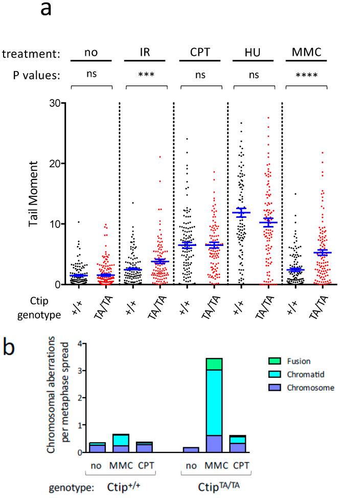
Homology-directed repair (HDR) of double-strand DNA breaks (DSBs) is dependent on enzymatic resection of DNA ends by the Mre11/Rad50/Nbs1 complex. DNA resection is triggered by the CtIP/Sae2 protein, which allosterically promotes Mre11-mediated endonuclease DNA cleavage at a position internal to the DSB. Although the mechanics of resection, including the initial endonucleolytic step, are largely conserved in eucaryotes, CtIP and its functional counterpart in Saccharomyces cerevisiae (Sae2) share only a modest stretch of amino acid homology. Nonetheless, this stretch contains two highly conserved phosphorylation sites for cyclin-dependent kinases (T843 in mouse) and the damage-induced ATM/ATR kinases (T855 in mouse), both of which are required for DNA resection. To explore the function of ATM/ATR phosphorylation at Ctip-T855, we generated and analyzed mice expressing the Ctip-T855A mutant. Surprisingly, unlike Ctip-null mice and Ctip-T843A-expressing mice, both of which undergo embryonic lethality, homozygous CtipT855A/T855A mice develop normally. Nonetheless, they are hypersensitive to ionizing radiation, and CtipT855A/T855A mouse embryo fibroblasts from these mice display marked defects in DNA resection, chromosomal stability, and HDR-mediated repair of DSBs. Thus, although ATM/ATR phosphorylation of CtIP-T855 is not required for normal animal development, it enhances CtIP-mediated DNA resection in response to acute stress, such as genotoxin exposure.

Keywords: ATM/ATR phosphorylation; CtIP; DNA break repair; DNA resection; Mre11; genotoxic stress; homologous recombination.


Conflict of interest statement

The authors declare no conflict of interest.

Figures

Figure 1
Figure 1
Homozygous CtipT855A/T855A mice and cells are hypersensitive to genotoxic stress. (a) Littermates of Ctip+/+, CtipT855A/+, and CtipT855A/T855A mice were subjected to whole-body irradiation (7.5 Gy) at five weeks of age and monitored for survival over the next three months. The number of mice in each genetic cohort is indicated. As determined by the log-rank (Mantel-Cox) test, the survival of CtipT855A/T855A mice was significantly reduced relative to their CtipT855A/+ (p = 0.04) and Ctip+/+ (p = 0.027) littermates. In contrast, the survival of CtipT855A/+ mice was statistically indistinguishable from that of Ctip+/+ mice (p = 0.74). (bd) Colony survival analysis of isogenic clones of Ctip+/+, CtipT855A/+, and CtipT855A/T855A mouse embryonic fibroblasts (MEFs) exposed to ionizing radiation (b), mitomycin C (c), or the PARP inhibitor olaparib (d). Survival was quantified as the percentage of colonies on genotoxin-treated cultures relative to untreated cultures. Each condition was tested in triplicate, and error bars represent SEM. For each culture, the Ctip genotype is indicated, and the specific MEF clone analyzed is denoted by the number in parentheses as listed in Table S1.
Figure 2
Figure 2
CtipT855A/T855A cells display elevated levels of DNA damage in response to specific genotoxic agents. (a) Levels of DNA damage in isogenic CtipT855A/T855A (clone 8) and Ctip+/+ (clone 7) MEFs were quantitated using the alkaline comet assay. Cells were either left untreated (no) or exposed to ionizing radiation (IR), camptothecin (CPT), hydroxyurea (HU), or mitomycin C (MMC). For each condition, the individual tail moments of at least 75 cells are presented as a dot plot; the mean tail moment is denoted by a horizontal blue line, and the SEM is indicated by error bars. p values as determined by the paired Student’s t test are non-significant (ns), <0.001 (***), or <0.0001 (****). (b) Chromosomal abnormalities were evaluated in isogenic CtipT855A/T855A (clone 8) and Ctip+/+ (clone 7) MEFs. Cells were left untreated (no) or exposed to either mitomycin C (MMC) or camptothecin (CPT), and the numbers of chromosome aberrations (fusion, chromatid-type, and chromosome-type) per metaphase spread were determined by telomere fluorescent in situ hybridization (T-FISH). (c) Representative T-FISH images of individual metaphase spreads, showing DAPI-stained chromosomes (blue) and Cy3/DAPI-stained telomeres (pink).
Figure 2
Figure 2
CtipT855A/T855A cells display elevated levels of DNA damage in response to specific genotoxic agents. (a) Levels of DNA damage in isogenic CtipT855A/T855A (clone 8) and Ctip+/+ (clone 7) MEFs were quantitated using the alkaline comet assay. Cells were either left untreated (no) or exposed to ionizing radiation (IR), camptothecin (CPT), hydroxyurea (HU), or mitomycin C (MMC). For each condition, the individual tail moments of at least 75 cells are presented as a dot plot; the mean tail moment is denoted by a horizontal blue line, and the SEM is indicated by error bars. p values as determined by the paired Student’s t test are non-significant (ns), <0.001 (***), or <0.0001 (****). (b) Chromosomal abnormalities were evaluated in isogenic CtipT855A/T855A (clone 8) and Ctip+/+ (clone 7) MEFs. Cells were left untreated (no) or exposed to either mitomycin C (MMC) or camptothecin (CPT), and the numbers of chromosome aberrations (fusion, chromatid-type, and chromosome-type) per metaphase spread were determined by telomere fluorescent in situ hybridization (T-FISH). (c) Representative T-FISH images of individual metaphase spreads, showing DAPI-stained chromosomes (blue) and Cy3/DAPI-stained telomeres (pink).
Figure 3
Figure 3
Homology-directed repair (HDR) of double-strand DNA breaks is impaired in CtipT855A/T855A cells. HDR efficiency was measured in independent subclones of Ctip+/+ (A, B, and C) and CtipT855A/T855A (D, E, and F) embryonic stem (ES) cells containing an integrated DR-GFP reporter. Cells were transfected with an empty (–) or I-SceI-expressing (I-SceI) vector, and the percentage of GFP-positive cells was quantified by flow cytometry. For comparison, isogenic ES cell subclones with (Brca1SF/SF) or without (Brca1+/+) a pathogenic mutation (S1598F) of the Brca1 tumor suppressor were evaluated in parallel. Each ES cell subclone was analyzed in triplicate, and error bars represent the SEM. Significant p values as determined by the paired Student’s t test are <0.05 (*), <0.01 (**), or <0.001 (***).
Figure 4
Figure 4
The formation of ssDNA/RPA filaments in response to DNA damage is impaired in CtipT855A/T855A cells. (a) Ctip+/+ (clone 7) and CtipT855A/T855A (clone 8) MEFs were exposed to camptothecin (CPT) for various time intervals (20, 60, and 90 min), and their cell lysates were immunoblotted with the indicated antibodies. (b) Homozygous mutant CtipT855A/T855A cells are defective in the formation of genotoxin-induced phospho-RPA2 foci. Ctip+/+ and CtipT855A/T855A MEFs, cultured with or without 1 mM camptothecin (CPT) for one hour, were evaluated by immunofluorescent microscopy for the presence of γH2AX-staining nuclear foci (left; indicative of DSBs) and phospho-RPA2-staining foci (right; indicative of ssDNA/RPA filaments). The histograms show the percentage of cells harboring >10 foci. For each culture, the Ctip genotype is indicated, and the specific MEF clone analyzed is denoted by the number in parentheses as listed in Table S1. The p values obtained using the paired Student’s t test were either non-specific (ns) or <0.01 (**). (c) Representative confocal images of isogenic CtipT855A/T855A (clone 8) and Ctip+/+ (clone 7) MEFs stained with DAPI (blue) and/or immunostained for phospho-RPA (T21) (green) and/or γH2AX (red). The white scale bars in the upper left images represent 20 µm.
Figure 5
Figure 5
SMART analysis of DNA resection in Ctip+/+, CtipT855A/T855A, and CtipS326A/S326A MEFs. (a) DNA fibers were prepared from pre-labeled (10 μM BrdU for 24 h) cell cultures that were left untreated (–) or exposed to either ionizing radiation (IR) or camptothecin (CPT). For each condition, the track lengths of at least 300 individual DNA fibers are presented as a dot plot, and the average track length is denoted by a horizontal bar. For each culture, the Ctip genotype is indicated, and the specific MEF clone analyzed (Table S1) is denoted in parentheses. The p values obtained using the Mann-Whitney rank sum test were either non-specific (ns) or <0.0001 (****). (b) SMART analysis of DNA resection in isogenic MEF clones expressing either wild-type Ctip (Ctip+/+) or a mutant (CtipS326A/S326A) impaired for interaction with Brca1 [38].

Similar articles

References

    1. Schaeper U., Subramanian T., Lim L., Boyd J.M., Chinnadurai G. Interaction between a cellular protein that binds to the C-terminal region of Adenovirus E1A (CtIP) and a novel cellular protein is disrupted by E1A through a conserved PLDLS motif. J. Biol. Chem. 1998;273:8549–8552. doi: 10.1074/jbc.273.15.8549. - DOI - PubMed
    1. Yu X., Wu L.C., Bowcock A.M., Aronheim A., Baer R. The carboxy-terminal (BRCT) motifs of BRCA interact in vivo with CtIP, a protein implicated in the CtBP pathway of transcriptional repression. J. Biol. Chem. 1998;273:25388–25392. doi: 10.1074/jbc.273.39.25388. - DOI - PubMed
    1. Wong A.K., Ormonde P.A., Pero R., Chen Y., Lian L., Salada G., Berry S., Lawrence Q., Dayananth P., Ha P., et al. Characterization of a carboxy-terminal BRCA1 interacting protein. Oncogene. 1998;17:2279–2285. doi: 10.1038/sj.onc.1202150. - DOI - PubMed
    1. Sartori A.A., Lukas C., Coates J., Fu S., Baer R., Lukas J., Jackson S.P. Human CtIP promotes DNA end resection. Nature. 2007;450:509–514. doi: 10.1038/nature06337. - DOI - PMC - PubMed
    1. Keeney S., Kleckner N. Covalent protein-DNA complexes at the 5’ strand termini of meiosis-specific double-strand breaks in yeast. Proc. Natl. Acad. Sci. USA. 1995;92:11274–11278. doi: 10.1073/pnas.92.24.11274. - DOI - PMC - PubMed

Publication types

LinkOut - more resources