. 2023 Nov 21;15(23):5500.
doi: 10.3390/cancers15235500.

Regulation of Vascular Endothelial Growth Factor Signaling by Nicotine in a Manner Dependent on Acetylcholine-and/or β-Adrenergic-Receptors in Human Lung Cancer Cells

Affiliations

Regulation of Vascular Endothelial Growth Factor Signaling by Nicotine in a Manner Dependent on Acetylcholine-and/or β-Adrenergic-Receptors in Human Lung Cancer Cells

Hind Al Khashali et al. Cancers (Basel). .

Abstract

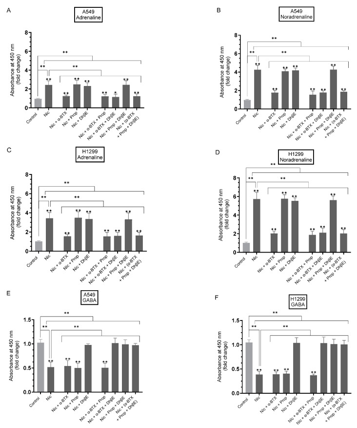
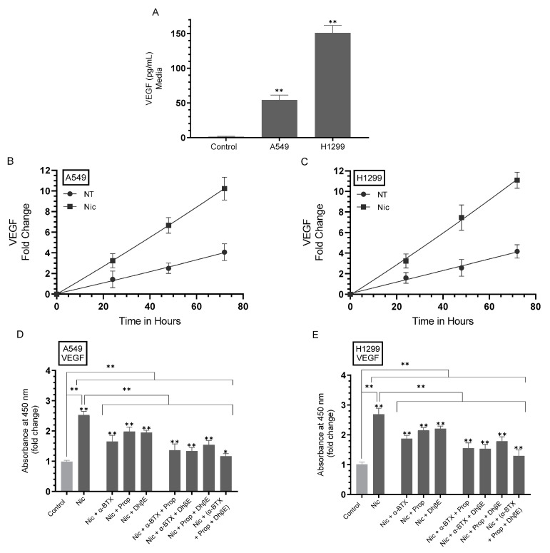
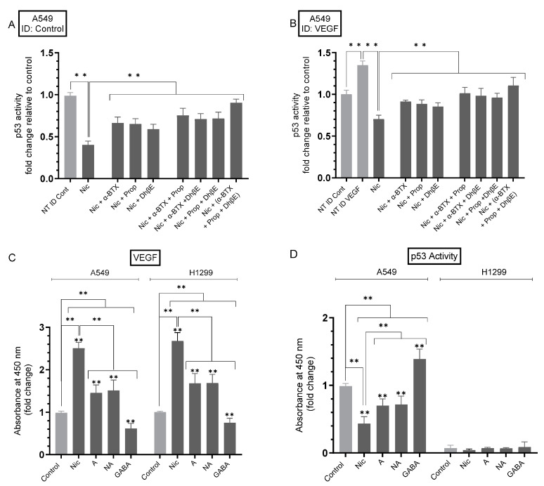
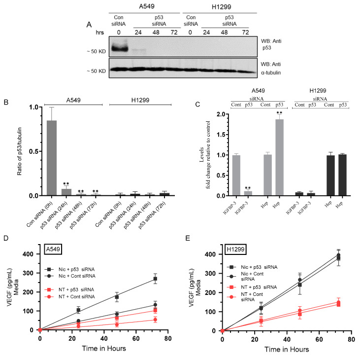
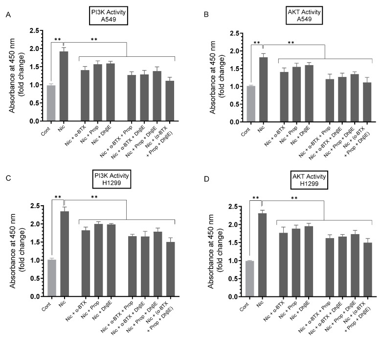
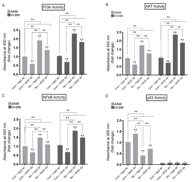
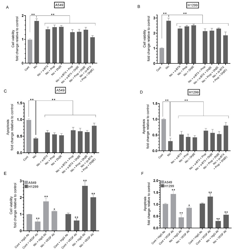
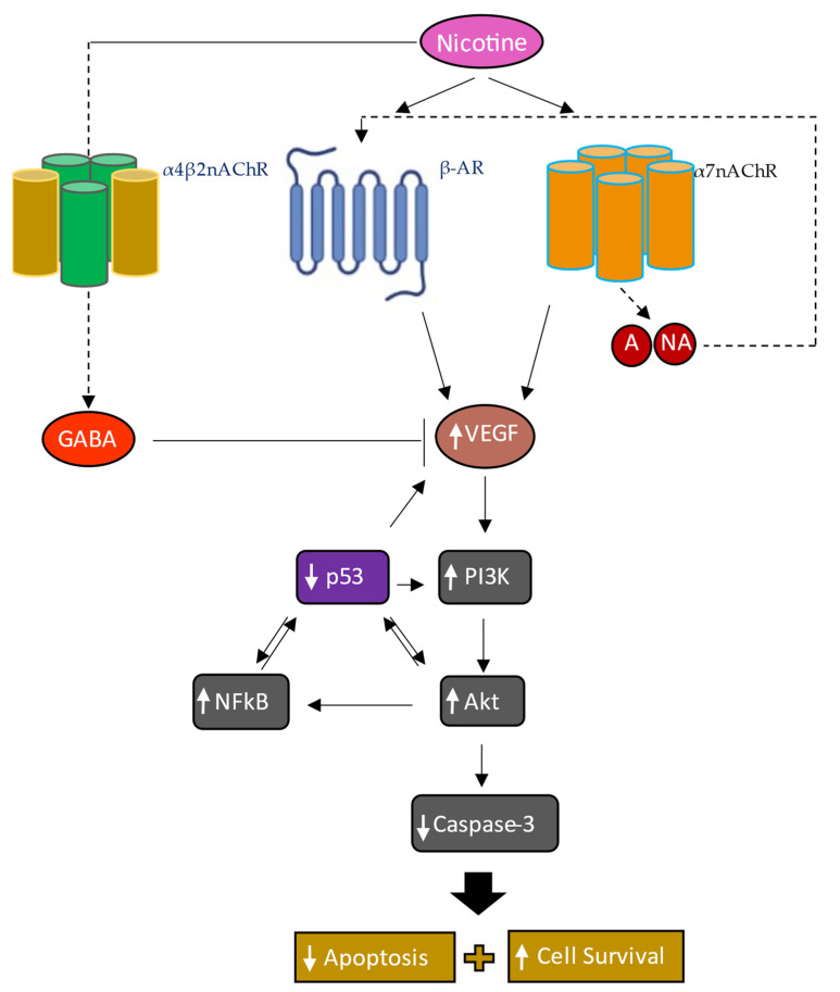
In addition to binding to nicotinic acetylcholine receptors (nAChRs), nicotine is known to regulate the β-adrenergic receptors (β-ARs) promoting oncogenic signaling. Using A549 (p53 wild-type) and H1299 (p53-null) lung cancer cells, we show that nicotine treatment led to: increased adrenaline/noradrenaline levels, an effect blocked by treatment with the α7nAChR inhibitor (α-BTX) but not by the β-blocker (propranolol) or the α4β2nAChR antagonist (DhβE); decreased GABA levels in A549 and H1299 cell media, an effect blocked by treatment with DhβE; increased VEGF levels and PI3K/AKT activities, an effect diminished by cell co-treatment with α-BTX, propranolol, and/or DhβE; and inhibited p53 activity in A549 cells, that was reversed, upon cell co-treatment with α-BTX, propranolol, and/or DhβE or by VEGF immunodepletion. VEGF levels increased upon cell treatment with nicotine, adrenaline/noradrenaline, and decreased with GABA treatment. On the other hand, the p53 activity decreased in A549 cells treated with nicotine, adrenaline/noradrenaline and increased upon cell incubation with GABA. Knockdown of p53 led to increased VEGF levels in the media of A549 cells. The addition of anti-VEGF antibodies to A549 and H1299 cells decreased cell viability and increased apoptosis; blocked the activities of PI3K, AKT, and NFκB in the absence or presence of nicotine; and resulted in increased p53 activation in A549 cells. We conclude that VEGF can be upregulated via α7nAChR and/or β-ARs and downregulated via GABA and/or p53 in response to the nicotine treatment of NSCLC cells.

Keywords: AKT; GABA; NFκB; PI3K; lung cancer; nicotine; nicotinic acetylcholine receptors; p53; vascular endothelial growth factor; β-adrenergic receptors.


Conflict of interest statement

The authors declare no conflict of interest.

Figures

Figure 1
Figure 1
The levels of adrenaline and noradrenaline increased in the media of A549 and H1299 cells treated with nicotine, while opposite effects were observed on GABA levels. Cells (0.2 × 10⁵) were grown in 10% FBS-supplemented media for 24 h. The following day, the cell monolayers were serum-starved for 24 h, then incubated in serum-free media for 72 h in the absence or presence of nicotine (Nic, 1 µM), α-bungarotoxin (α-BTX, 200 nM), propranolol (Prop, 1 µM), DhβE (10 μM), or in combination. The concentrations of adrenaline (A,C), noradrenaline (B,D), and GABA (E,F) were measured as described in Section 2. Data were expressed as a fold change relative to the control untreated cells (Control) using the GraphPad 9.5.1 software (n = 5). Asterisks indicate a statistically significant difference from the corresponding control for each cell line. Statistical differences between different groups were analyzed by an ordinary one-way analysis of variance (ANOVA) followed by Tukey’s post hoc multiple comparison test, * p < 0.05, ** p < 0.01.
Figure 2
Figure 2
The levels of VEGF increased in the media of A549 and H1299 cells upon treatment with nicotine, an effect blocked by cell co-treatment with α-BTX, propranolol, and/or DhβE. Cells (0.2 × 10⁵) were grown in 10% FBS-supplemented media for 24 h. The following day, the cell monolayers were serum-starved for 24 h, the media replaced with fresh serum-free media (0 h) then the cells incubated for 72 h (A), and the concentration of VEGF was measured as described in Section 2. Control was media not incubated with cells. (B,C) The levels of VEGF were measured as a function of time without, or with, nicotine (Nic, 1 µM) and expressed as a fold change relative to 0 h. (D,E) The levels of VEGF in the presence of Nic without, or with, α-bungarotoxin (α-BTX, 200 nM), propranolol (Prop, 1 µM), DhβE (10 μM), or in combination were determined after 72 h of incubation. Data were averaged and expressed as a fold change relative to the control using the GraphPad 9.5.1 software (n = 5). Asterisks indicate a statistically significant difference from the corresponding controls while absence of asterisks indicates no significance. Statistical differences between different groups were analyzed by ANOVA followed by Tukey’s post hoc multiple comparison test, * p < 0.05, ** p < 0.01.
Figure 3
Figure 3
The activity of p53 is inhibited upon A549 cell treatment with nicotine, an effect partially reversed upon cell co-treatment with α-BTX, propranolol, and/or DhβE or by using media immunodepleted (ID) of VEGF. In addition, the levels of VEGF increased in the media of A549 and H1299 cells treated with nicotine, adrenaline, or noradrenaline and decreased by cell treatment with GABA while opposite effects were found on the p53 activity in A549 cells. Cells (0.2 × 10⁵) were grown in 10% FBS-supplemented media for 24 h. The following day, the cell monolayers were serum-starved for 24 h, then incubated in serum-free media for 72 h in the absence or presence of nicotine (Nic, 1 µM), α-bungarotoxin (α-BTX, 200 nM), propranolol (Prop, 1 µM), DhβE (10 μM), adrenaline (A, 10 nM), noradrenaline (NA, 10 nM), GABA (10 µM), or in combination. The activity of p53 was measured using cells incubated with media collected and ID using hIgG (20 μg/mL) as a control (A) or anti-VEGF-specific antibodies (B) (20 μg/mL) (Section 2). The concentration of VEGF (C) and the p53 activity (D) were measured (Section 2). Data were expressed as a fold change relative to cells not treated (NT) and ID with hIgG control (NT ID Cont) (A,B), control untreated cells (Control) of each cell line (C) or to A549 control (D) using the GraphPad 9.5.1 software (n = 5). Asterisks indicate a statistically significant difference while absence of asterisks indicates no significance. Statistical differences between different groups were analyzed by ANOVA followed by Tukey’s post hoc multiple comparison test, ** p < 0.01.
Figure 4
Figure 4
Knockdown of p53 increased the levels of VEGF in the media of A549 cells untreated or treated with nicotine. Cells (0.2 × 10⁵) were grown in media supplemented with 10% FBS overnight. The cell monolayers were then incubated in serum-free media for 24 h, then the media were replaced with fresh serum-free media (0 h). The cells were then incubated with the indicated siRNAs and either not treated (NT) or treated with nicotine (Nic, 1 µM) (Section 2). Western blotting (A) using the indicated antibodies was carried out on the same concentration of total protein (15 µL of 600 µg/mL) of the cell lysates. Quantitation of the Western blot, performed three times, was carried out using Image J 1.47 v software (B). To further verify p53 knockdown, the levels of IGFBP-3 and heparanase (Hep) were measured (C). The media were used to quantitate the levels of VEGF (Section 2, (D,E), n = 5), using the same concentration of protein (3 µL of 600 µg/mL total protein), as a function of time. Data were plotted using the GraphPad 9.5.1 software, ** p < 0.01.
Figure 5
Figure 5
The activities of PI3K and AKT were increased upon A549 and H1299 cell treatment with nicotine (Nic) and decreased upon cell co-treatment with Nic and α-BTX, propranolol, and/or DhβE. Cells (0.2 × 10⁵) were grown in media supplemented with 10% FBS overnight. Cells were then serum-starved for 24 h, then incubated in serum-free media for 72 h in the absence or presence of nicotine (Nic, 1 µM), α-bungarotoxin (α-BTX, 200 nM), propranolol (Prop, 1 µM), DhβE (10 μM), or in combination. The PI3K activities (A,C) and AKT activities (B,D) were measured (Section 2). Data were expressed as a fold change relative to the control untreated cells (Control) using the GraphPad 9.5.1 software (n = 5). Statistical differences between different groups were analyzed by ANOVA followed by Tukey’s post hoc multiple comparison test, ** p < 0.01.
Figure 6
Figure 6
Anti-VEGF antibodies decreased the activities of PI3K, AKT, and NFκB in A549 and H1299 cells without or with nicotine and increased p53 activation in A549 cells. Cells were grown in media with 10% FBS for 24 h, serum-starved overnight, then incubated in serum-free media for 72 h without or with nicotine (Nic, 1 µM), hIgG (20 μg/mL) as a control, anti-VEGF-specific antibodies (20 μg/mL), or in combination. The activities of PI3K, AKT, NFκB, and p53 were measured (Section 2). Data were averaged and expressed as a fold change relative to the control cells treated with hIgG (Cont + hIgG Ab) of each cell line (AC) or to A549 (Cont + hIgG Ab) (D) using the GraphPad 9.5.1 software (n = 5). Asterisks indicate a statistically significant difference from the control of each cell line. Statistical differences between different groups were analyzed by an ordinary one-way analysis of variance (ANOVA) followed by Tukey’s post hoc multiple comparison test, ** p < 0.01.
Figure 7
Figure 7
Compared to A549 and H1299 cell treatment with only nicotine (Nic), co-treatment with Nic and either α-BTX, propranolol, DhβE, or in combination resulted in decreased cell viability and increased apoptosis. In addition, anti-VEGF antibodies decreased cell viability and increased apoptosis of A549 and H1299 cells without, or with, nicotine treatment. Cells were grown in media with 10% FBS for 24 h, serum-starved overnight, then incubated in serum-free media for 72 h in the absence or presence of nicotine (Nic, 1 µM), α-bungarotoxin (α-BTX, 200 nM), propranolol (Prop, 1 µM), DhβE (10 μM), hIgG (20 μg/mL) as a control or anti-VEGF-specific antibodies (20 μg/mL), or in combination. Cell viability (A,B,E) and apoptosis (C,D,F) were measured (Section 2). Data were averaged, normalized, and expressed as a fold change relative to the control untreated cells (Cont) (AD) or to control cells treated with hIgG (Cont + hIgG Ab) of each cell line (E,F) using the GraphPad 9.5.1 software (n = 5). Statistical differences between different groups were analyzed by an ordinary one-way analysis of variance (ANOVA) followed by Tukey’s post hoc multiple comparison test, * p < 0.05, ** p < 0.01.
Figure 8
Figure 8
Summary of the main findings of this study. Nicotine enhances VEGF signaling in NSCLC by acting positively via the α7nAChR and β-ARs and negatively regulating GABA levels. Nicotine treatment leads to increased levels of adrenaline (A), noradrenaline (NA), VEGF, and activities of PI3K, AKT, NFκB, and decreased p53 activity via a mechanism involving α7nAChR, α4β2nAChR, and β-ARs leading to increased cell survival and decreased apoptosis. Dashed lines indicate a potential indirect effect. Colored boxes indicate new mechanisms provided from this study in NSCLC cells while gray boxes show findings from previous studies, from our laboratory and others, repeated here in this study under the same conditions to provide the overall scheme. While nicotine treatment led to increased levels of A/NA and decreased GABA levels in A549 and H1299 cell media, further evidence is needed to establish the direct involvement of the α7nAChR and α4β2nAChR in the regulation of these levels. Similarly, further experiments are needed to examine whether p53 acts directly on VEGF and vice versa.

Similar articles

Cited by

References

    1. Siegel R.L., Miller K.D., Jemal A. Cancer Statistics, 2016. CA Cancer J. Clin. 2016;66:7–30. doi: 10.3322/caac.21332. - DOI - PubMed
    1. Siegel R., Ma J., Zou Z., Jemal A. Cancer Statistics, 2014. CA Cancer J. Clin. 2014;64:9–29. doi: 10.3322/caac.21208. - DOI - PubMed
    1. Rotow J., Bivona T.G. Understanding and Targeting Resistance Mechanisms in NSCLC. Nat. Rev. Cancer. 2017;17:637–658. doi: 10.1038/nrc.2017.84. - DOI - PubMed
    1. Siddiqui F., Siddiqui A.H. StatPearls. StatPearls Publishing; Treasure Island, FL, USA: 2021. Lung Cancer.
    1. Chen R.-J., Chang L.W., Lin P., Wang Y.-J. Epigenetic Effects and Molecular Mechanisms of Tumorigenesis Induced by Cigarette Smoke: An Overview. J. Oncol. 2011;2011:654931. doi: 10.1155/2011/654931. - DOI - PMC - PubMed