

Melanin-The Éminence Grise of Melanoma and Parkinson's Disease Development
- PMID: 38067245
- PMCID: PMC10705212
- DOI: 10.3390/cancers15235541
Melanin-The Éminence Grise of Melanoma and Parkinson's Disease Development
Abstract
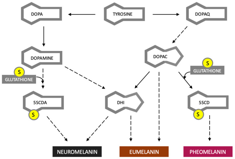
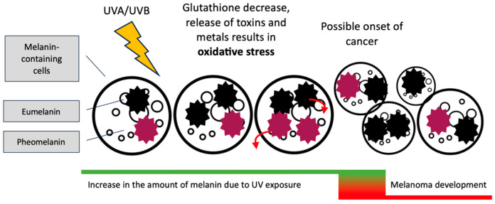
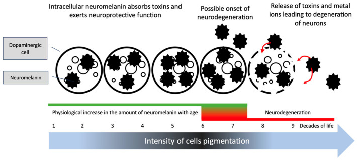
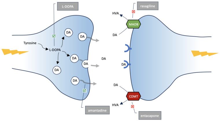
A common feature of Parkinson's disease (PD) and melanoma is their starting points being based on cells capable of converting tyrosine into melanin. Melanocytes produce two types of melanin: eumelanin and pheomelanin. These dyes are designed to protect epidermal cells from the harmful effects of UV radiation. Neurones of the substantia nigra, which degenerate during PD, produce neuromelanin, the physiological role of which is not fully explained. This article discusses the potential role of melanins in the pathogenesis of both diseases. Melanins, due to their ability to accumulate toxic substances, may become their sources over time. The use of glutathione for the synthesis of pheomelanins and neuromelanins may reduce the antioxidant capacity of cells, leading to an excessive synthesis of free radicals. This study also tested the hypothesis that certain drugs used in the treatment of PD (L-DOPA, MAO-B and COMT inhibitors, and amantadine), aimed at increasing dopamine concentration, could potentially contribute to the development of melanoma. The role and properties of melanins should continue to be researched. Whether excessive melanin synthesis or its accumulation in the extracellular space may be factors initiating the development of diseases remains an open question.
Keywords: Parkinson’s disease; dopamine; eumelanin; melanoma; neuromelanin; pheomelanin.
Conflict of interest statement
The authors declare no conflict of interest.
Figures
Similar articles
-
DOPA pheomelanin is increased in nigral neuromelanin of Parkinson's disease.Prog Neurobiol. 2023 Apr;223:102414. doi: 10.1016/j.pneurobio.2023.102414. Epub 2023 Feb 4. Prog Neurobiol. 2023. PMID: 36746222
-
Neuromelanins in brain aging and Parkinson's disease: synthesis, structure, neuroinflammatory, and neurodegenerative role.IUBMB Life. 2023 Jan;75(1):55-65. doi: 10.1002/iub.2654. Epub 2022 Jun 11. IUBMB Life. 2023. PMID: 35689524 Free PMC article.
-
Melanin and Neuromelanin Fluorescence Studies Focusing on Parkinson's Disease and Its Inherent Risk for Melanoma.Cells. 2019 Jun 15;8(6):592. doi: 10.3390/cells8060592. Cells. 2019. PMID: 31208049 Free PMC article.
-
The enigma of neuromelanin in Parkinson's disease substantia nigra.J Neural Transm Suppl. 1994;43:113-22. J Neural Transm Suppl. 1994. PMID: 7884393 Review.
-
Neuromelanin in Parkinson's Disease: Tyrosine Hydroxylase and Tyrosinase.Int J Mol Sci. 2022 Apr 10;23(8):4176. doi: 10.3390/ijms23084176. Int J Mol Sci. 2022. PMID: 35456994 Free PMC article. Review.
Cited by
-
Anti-Melanogenic Effects of Takifugu flavidus Muscle Hydrolysate in B16F10 Melanoma Cells and Zebrafish.Mar Drugs. 2024 Apr 29;22(5):206. doi: 10.3390/md22050206. Mar Drugs. 2024. PMID: 38786597 Free PMC article.
-
The Invisible Fraction within Melanin Capable of Absorbing UV Light and with Fluorescent Properties: Is It Lacking Consideration?Int J Mol Sci. 2024 Aug 3;25(15):8490. doi: 10.3390/ijms25158490. Int J Mol Sci. 2024. PMID: 39126061 Free PMC article.
-
Secretory pathway in honeybee venom and actin: evidence from chemical-biological interactions.Hum Cell. 2025 May 23;38(4):104. doi: 10.1007/s13577-025-01240-9. Hum Cell. 2025. PMID: 40410651 No abstract available.
References
-
- Rastrelli M., Tropea S., Rossi C.R., Alaibac M. Melanoma: Epidemiology, Risk Factors, Pathogenesis, Diagnosis and Classification. [(accessed on 14 November 2014)];In Vivo. 2014 28:1005–1011. Available online: http://iv.iiarjournals.org/content/28/6/1005.abstract. - PubMed
Publication types
LinkOut - more resources
Full Text Sources
Miscellaneous
