. 2023 Nov 24;15(23):5571.
doi: 10.3390/cancers15235571.

Identification of Tumor-Suppressive miR-139-3p- Regulated Genes: TRIP13 as a Therapeutic Target in Lung Adenocarcinoma

Affiliations

Identification of Tumor-Suppressive miR-139-3p- Regulated Genes: TRIP13 as a Therapeutic Target in Lung Adenocarcinoma

Yoko Hagihara et al. Cancers (Basel). .

Abstract

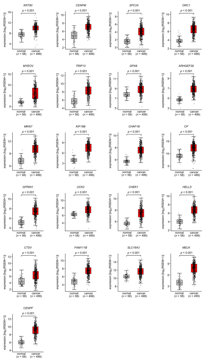
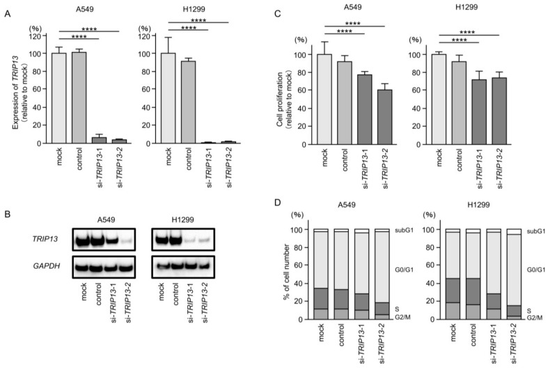
Analyses of our microRNA (miRNA) expression signature combined with The Cancer Genome Atlas (TCGA) data revealed that both strands of pre-miR-139 (miR-139-5p, the guide strand, and miR-139-3p, the passenger strand) are significantly downregulated in lung adenocarcinoma (LUAD) clinical specimens. Functional analyses of LUAD cells ectopically expressing miR-139-3p showed significant suppression of their aggressiveness (e.g., cancer cell proliferation, migration, and invasion). The involvement of the passenger strand, miR-139-3p, in LUAD pathogenesis, is an interesting finding contributing to the elucidation of unknown molecular networks in LUAD. Of 1108 genes identified as miR-139-3p targets in LUAD cells, 21 were significantly upregulated in LUAD tissues according to TCGA analysis, and their high expression negatively affected the prognosis of LUAD patients. We focused on thyroid hormone receptor interactor 13 (TRIP13) and investigated its cancer-promoting functions in LUAD cells. Luciferase assays showed that miR-139-3p directly regulated TRIP13. siRNA-mediated TRIP13 knockdown and TRIP13 inhibition by a specific inhibitor (DCZ0415) attenuated the malignant transformation of LUAD cells. Interestingly, when used in combination with anticancer drugs (cisplatin and carboplatin), DCZ0415 exerted synergistic effects on cell proliferation suppression. Identifying the molecular pathways regulated by tumor-suppressive miRNAs (including passenger strands) may aid in the discovery of diagnostic markers and therapeutic targets for LUAD.

Keywords: TRIP13; lung adenocarcinoma; miR-139-3p; microRNA; passenger strand.


Conflict of interest statement

The authors declare no conflict of interest.

Figures

Figure 1
Figure 1
Expression levels of miR-139-5p and miR-139-3p in LUAD clinical tissues. (A) Volcano plot of the miRNA expression signature based on miRNA sequencing (GEO accession number: GSE230229). The log2 fold change (FC) in expression is plotted on the x-axis and the log10 p-value on the y-axis. The blue and red dots represent the downregulated (log2FC < −2.0 and p < 0.05) and upregulated (log2FC > 2.0 and p < 0.05) miRNAs, respectively. (B) Chromosomal location of pre-miR-139 within the human genome. The mature sequences of miR-139-5p (guide strand) and miR-139-3p (passenger strand) are shown. (C) Expression levels of miR-139-5p and miR-139-3p validated in LUAD clinical specimens. The expression of both miRNAs was significantly downregulated in cancer tissues (p < 0.001). (D) Positive correlations (Spearman’s rank test) between miR-139-5p and miR-139-3p expression levels in clinical specimens (r = 0.513, p < 0.001).
Figure 2
Figure 2
Effects of ectopic expression of miR-139-3p in LUAD cells (A549 and H1299). (A) Cell proliferation assessed by XTT assay. At 72 h after transient transfection of miRNAs, cancer cell viability was analyzed. (B) Cell cycle status at 72 h after transfection with miR-139-3p assessed using flow cytometry. (C) Cell invasion assessed using Matrigel invasion assays at 48 h after seeding miR-139-3p-transfected cells into the chambers. (D) Cell migration assessed using a membrane culture system at 48 h after seeding miR-139-3p transfected cells into the chambers. ****, p < 0.0001.
Figure 3
Figure 3
Flowchart for identification of miR-139-3p targets in LUAD cell. To identify putative targets of miR-139-3p in LUAD cells, the following two datasets were merged: the TargetScanHuman database (release 8.0) and our original mRNA expression profile (miR-139-3p-transfected A549 cells; GEO accession number: GSE242241). A total of 1108 genes were identified as putative miR-139-3p targets. Furthermore, we searched for genes that were associated with the prognosis of LUAD patients using two databases: GEPIA (http://gepia2.cancer-pku.cn/#analysis (accessed on 17 January 2023)) and OncoLnc (http://www.oncolnc.org (accessed on 17 January 2023)). Of the miR-139-3p target genes, 21 were upregulated in LUAD tissues, and these 21 genes were analyzed further.
Figure 4
Figure 4
Expression levels of the 21 target genes regulated by miR-139-3p in LUAD. The expression levels of the 21 miR-139-3p target genes (KRT80, CENPM, SPC24, ORC1, MYEOV, TRIP13, GPX8, ARHGEF39, MKI67, KIF18B, CHAF1B, CP, GPRIN1, UCK2, CHEK1, HELLS, CTSV, FAM111B, SLC16A3, MELK, and CENPF) in LUAD clinical specimens were analyzed using the TCGA-LUAD dataset. All genes were upregulated in LUAD tissues (n = 499) compared with normal tissues (n = 58) (p < 0.001).
Figure 5
Figure 5
Gene expression in, and 5-year overall survival rate of, patients with LUAD. Kaplan–Meier curves of the 5-year overall survival rates according to the expression of the 21 target genes (KRT80, CENPM, SPC24, ORC1, MYEOV, TRIP13, GPX8, ARHGEF39, MKI67, KIF18B, CHAF1B, CP, GPRIN1, UCK2, CHEK1, HELLS, CTSV, FAM111B, SLC16A3, MELK, and CENPF) are shown. Low expression of all 21 genes was significantly predictive of poorer overall survival in patients with LUAD. The patients (n = 487) were divided into high- and low-expression groups according to the median gene expression level. The red and blue lines represent the high and low expression groups, respectively.
Figure 6
Figure 6
Direct regulation of TRIP13 by miR-139-3p expression in LUAD cells. (A) Significant reduction in the TRIP13 mRNA level by ectopic expression of miR-139-3p in LUAD cells (A549 and H1299). Total RNA was extracted 72 h after miR-139-3p transfection into LUAD cells, and expression levels were analyzed by real-time PCR. For miRNA expression, GAPDH was used as an internal control. (B) Significant reduction in the TRIP13 protein level by ectopic expression of miR-139-3p in LUAD cells (A549 and H1299). Protein level expression was determined by Western blotting. Proteins were collected 72 h after miR-139-3p transfection. GAPDH was used as an internal control. (C) Putative miR-139-3p binding sites in the 3′UTR of the TRIP13 gene detected in the TargetScanHuman database (release 8.0). (D) Direct binding of miR-139-3p to target sequences was analyzed by dual luciferase reporter assays. These data showed that miR-139-3p bound directly to the target sequence. ***, p < 0.001; ****, p < 0.0001; N.S., not significant.
Figure 7
Figure 7
Clinical significance of TRIP13 expression in LUAD. (A) Immunohistochemical staining of TRIP13. Immunostaining showed that the TRIP13 protein was strongly expressed in cancer lesions and less in non-cancerous tissues. Scale bar: 200 µm (low magnification); 50 µm (high magnification). (B) Forest plot showing the results of multivariate Cox proportional hazards regression analysis of the 5-year overall survival rate. Patients with high TRIP13 expression had a significantly lower overall survival rate. These data were obtained from TCGA-LUAD datasets. (C) Gene set enrichment analysis (GSEA) was applied to explore molecular pathways mediated by TRIP13 in LUAD cells. The top six pathways enriched in LUAD patients with high TRIP13 expression were cell cycle, DNA replication, proteasome, P53 signaling pathway, homologous recombination, and mismatch repair.
Figure 7
Figure 7
Clinical significance of TRIP13 expression in LUAD. (A) Immunohistochemical staining of TRIP13. Immunostaining showed that the TRIP13 protein was strongly expressed in cancer lesions and less in non-cancerous tissues. Scale bar: 200 µm (low magnification); 50 µm (high magnification). (B) Forest plot showing the results of multivariate Cox proportional hazards regression analysis of the 5-year overall survival rate. Patients with high TRIP13 expression had a significantly lower overall survival rate. These data were obtained from TCGA-LUAD datasets. (C) Gene set enrichment analysis (GSEA) was applied to explore molecular pathways mediated by TRIP13 in LUAD cells. The top six pathways enriched in LUAD patients with high TRIP13 expression were cell cycle, DNA replication, proteasome, P53 signaling pathway, homologous recombination, and mismatch repair.
Figure 8
Figure 8
Effects of knockdown of TRIP13 by siRNAs in LUAD cells (A549 and H1299). (A) TRIP13 mRNA levels were effectively blocked by each siRNA in LUAD cells (A549 and H1299). (B) TRIP13 protein levels were effectively inhibited by two siRNAs (siTRIP13-1 and siTRIP13-2) in LUAD cells (A549 and H1299). (C) Cell proliferation was assessed using XTT assays 72 h after siRNA transfection into LUAD cells. Cell proliferation was significantly blocked after transient transfection of siRNAs. (D) Flow cytometry analysis of cell cycle status 72 h after transfection with siTRIP13-1 and siTRIP13-2. ****, p < 0.0001.
Figure 9
Figure 9
Effects of treatment with DCZ0415 (TRIP13 inhibitor) on LUAD cells (A549 and H1299). The proliferation of LUAD cells was significantly inhibited by DCZ0415 in a concentration-dependent manner. ****, p < 0.0001; N.S., not significant.
Figure 10
Figure 10
Effects of co-treatment of DCZ0415 (TRIP13 inhibitor) with anticancer drugs (cisplatin and carboplatin) in LUAD cells (A549 and H1299). LUAD cells showed increased sensitivity to anticancer drugs (cisplatin and carboplatin) when co-treated with DCZ0415. CDDP, cisplatin; CBDCA, carboplatin.
Figure 11
Figure 11
Synergistic effects between two anticancer drugs (cisplatin and carboplatin) and the TRIP13 inhibitor DCZ0415. The Chou–Talalay method was used to determine the synergistic effects between two anticancer drugs (cisplatin and carboplatin) and DCZ0415 in LUAD cells (A549 and H1299). CDDP, cisplatin; CBDCA, carboplatin.

Similar articles

Cited by

References

    1. Sung H., Ferlay J., Siegel R.L., Laversanne M., Soerjomataram I., Jemal A., Bray F. Global Cancer Statistics 2020: GLO-BOCAN Estimates of Incidence and Mortality Worldwide for 36 Cancers in 185 Countries. CA Cancer J. Clin. 2021;71:209–249. doi: 10.3322/caac.21660. - DOI - PubMed
    1. Siegel R.L., Miller K.D., Fuchs H.E., Jemal A. Cancer statistics, 2022. CA Cancer J. Clin. 2022;72:7–33. doi: 10.3322/caac.21708. - DOI - PubMed
    1. Thai A.A., Solomon B.J., Sequist L.V., Gainor J.F., Heist R.S. Lung cancer. Lancet. 2021;398:535–554. - PubMed
    1. Alexander M., Kim S.Y., Cheng H. Update 2020: Management of Non-Small Cell Lung Cancer. Lung. 2020;198:897–907. doi: 10.1007/s00408-020-00407-5. - DOI - PMC - PubMed
    1. Fatica A., Bozzoni I. Long non-coding RNAs: New players in cell differentiation and development. Nat. Rev. Genet. 2014;15:7–21. doi: 10.1038/nrg3606. - DOI - PubMed