

Is System xc- a Suitable Target for Tumour Detection and Response Assessment with Imaging?
- PMID: 38067277
- PMCID: PMC10705217
- DOI: 10.3390/cancers15235573
Is System xc- a Suitable Target for Tumour Detection and Response Assessment with Imaging?
Abstract
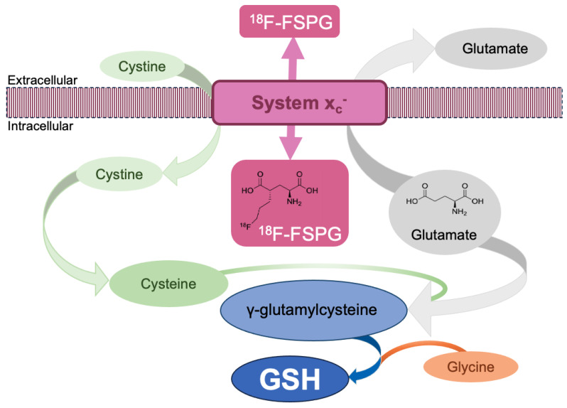
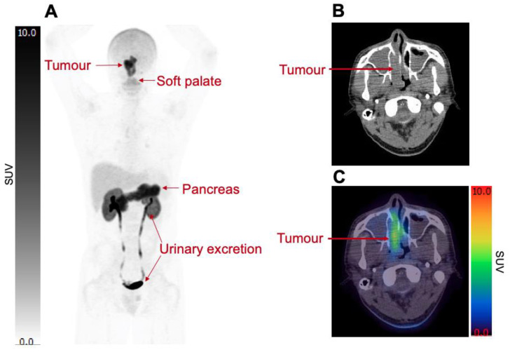
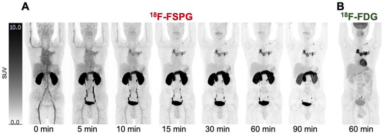
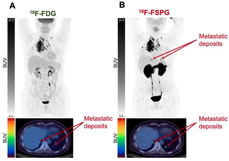
System xc- is upregulated in cancer cells and can be imaged using novel radiotracers, most commonly with (4S)-4-(3-[18F]fluoropropyl)-L-glutamic acid (18F-FSPG). The aim of this review was to summarise the use of 18F-FSPG in humans, explore the benefits and limitations of 18F-FSPG, and assess the potential for further use of 18F-FSPG in cancer patients. To date, ten papers have described the use of 18F-FSPG in human cancers. These studies involved small numbers of patients (range 1-26) and assessed the use of 18F-FSPG as a general oncological diagnostic agent across different cancer types. These clinical trials were contrasting in their findings, limiting the scope of 18F-FSPG PET/CT as a purely diagnostic agent, primarily due to heterogeneity of 18F-FSPG retention both between cancer types and patients. Despite these limitations, a potential further application for 18F-FSPG is in the assessment of early treatment response and prediction of treatment resistance. Animal models of cancer have shown that changes in 18F-FSPG retention following effective therapy precede glycolytic changes, as indicated by 18F-FDG, and changes in tumour volume, as measured by CT. If these results could be replicated in human clinical trials, imaging with 18F-FSPG PET/CT would offer an exciting route towards addressing the currently unmet clinical needs of treatment resistance prediction and early imaging assessment of therapy response.
Keywords: redox status; response assessment; system xc−; treatment resistance; treatment response.
Conflict of interest statement
The authors declare no conflicts of interest.
Figures
Similar articles
-
Clinical [18F]FSPG Positron Emission Tomography Imaging Reveals Heterogeneity in Tumor-Associated System xc- Activity.Cancers (Basel). 2024 Apr 8;16(7):1437. doi: 10.3390/cancers16071437. Cancers (Basel). 2024. PMID: 38611114 Free PMC article.
-
Radiotracer stereochemistry affects substrate affinity and kinetics for improved imaging of system xC- in tumors.Theranostics. 2022 Jan 24;12(4):1921-1936. doi: 10.7150/thno.63237. eCollection 2022. Theranostics. 2022. PMID: 35198080 Free PMC article.
-
Initial evaluation of (4S)-4-(3-[18F]fluoropropyl)-L-glutamate (FSPG) PET/CT imaging in patients with head and neck cancer, colorectal cancer, or non-Hodgkin lymphoma.EJNMMI Res. 2020 Aug 28;10(1):100. doi: 10.1186/s13550-020-00678-2. EJNMMI Res. 2020. PMID: 32857284 Free PMC article.
-
(4S)-4-(3-[18F]Fluoropropyl)-l-glutamate.2013 Mar 20 [updated 2013 May 23]. In: Molecular Imaging and Contrast Agent Database (MICAD) [Internet]. Bethesda (MD): National Center for Biotechnology Information (US); 2004–2013. 2013 Mar 20 [updated 2013 May 23]. In: Molecular Imaging and Contrast Agent Database (MICAD) [Internet]. Bethesda (MD): National Center for Biotechnology Information (US); 2004–2013. PMID: 23700646 Free Books & Documents. Review.
-
The lung cancers: staging and response, CT, 18F-FDG PET/CT, MRI, DWI: review and new perspectives.Br J Radiol. 2023 Aug;96(1148):20220339. doi: 10.1259/bjr.20220339. Epub 2023 May 17. Br J Radiol. 2023. PMID: 37097296 Free PMC article. Review.
Cited by
-
Novel molecular imaging approaches in oncology: towards a more accurate estimation of tumour response.Curr Opin Oncol. 2025 Sep 1;37(5):522-528. doi: 10.1097/CCO.0000000000001166. Epub 2025 Jul 9. Curr Opin Oncol. 2025. PMID: 40637055 Free PMC article. Review.
-
[18F]FSPG-PET provides an early marker of radiotherapy response in head and neck squamous cell cancer.Npj Imaging. 2024;2(1):28. doi: 10.1038/s44303-024-00038-y. Epub 2024 Aug 9. Npj Imaging. 2024. PMID: 39132311 Free PMC article.
-
18F-5-fluoro-aminosuberic acid PET/CT imaging of oxidative-stress features during the formation of DEN-induced rat hepatocellular carcinoma.Theranostics. 2025 Jan 1;15(1):141-154. doi: 10.7150/thno.100467. eCollection 2025. Theranostics. 2025. PMID: 39744233 Free PMC article.
-
Clinical [18F]FSPG Positron Emission Tomography Imaging Reveals Heterogeneity in Tumor-Associated System xc- Activity.Cancers (Basel). 2024 Apr 8;16(7):1437. doi: 10.3390/cancers16071437. Cancers (Basel). 2024. PMID: 38611114 Free PMC article.
-
PET Imaging of Metabolism, Perfusion, and Hypoxia: FDG and Beyond.Cancer J. 2024 May-Jun 01;30(3):159-169. doi: 10.1097/PPO.0000000000000716. Cancer J. 2024. PMID: 38753750 Free PMC article. Review.
References
-
- McCormick P.N., Greenwood H.E., Glaser M., Maddocks O.D.K., Gendron T., Sander K., Gowrishankar G., Hoehne A., Zhang T., Shuhendler A.J., et al. Assessment of Tumor Redox Status through (S)-4-(3-[18F]Fluoropropyl)-l-Glutamic Acid Positron Emission Tomography Imaging of System Xc- Activity. Cancer Res. 2018;79:853–863. doi: 10.1158/0008-5472.CAN-18-2634. - DOI - PMC - PubMed
Publication types
Grants and funding
LinkOut - more resources
Full Text Sources
