The Role of Gene Fusions in Thymic Epithelial Tumors
- PMID: 38067300
- PMCID: PMC10705505
- DOI: 10.3390/cancers15235596
The Role of Gene Fusions in Thymic Epithelial Tumors
Abstract
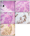
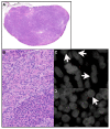
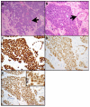
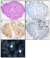
Thymic epithelial tumors (TET) are rare and large molecular studies are therefore difficult to perform. However, institutional case series and rare multi-institutional studies have identified a number of interesting molecular aberrations in TET, including gene fusions in a subset of these tumors. These gene fusions can aid in the diagnosis, shed light on the pathogenesis of a subset of tumors, and potentially may provide patients with the opportunity to undergo targeted therapy or participation in clinical trials. Gene fusions that have been identified in TET include MAML2 rearrangements in 50% to 56% of mucoepidermoid carcinomas (MAML2::CRTC1), 77% to 100% of metaplastic thymomas (YAP1::MAML2), and 6% of B2 and B3 thymomas (MAML2::KMT2A); NUTM1 rearrangements in NUT carcinomas (most commonly BRD4::NUTM1); EWSR1 rearrangement in hyalinizing clear cell carcinoma (EWSR1::ATF1); and NTRK rearrangement in a thymoma (EIF4B::NTRK3). This review focuses on TET in which these fusion genes have been identified, their morphologic, immunophenotypic, and clinical characteristics and potential clinical implications of the fusion genes. Larger, multi-institutional, global studies are needed to further elucidate the molecular characteristics of these rare but sometimes very aggressive tumors in order to optimize patient management, provide patients with the opportunity to undergo targeted therapy and participate in clinical trials, and to elucidate the pathogenesis of these tumors.
Keywords: EWSR1; MAML2; NTRK; NUTM1; YAP1.
Conflict of interest statement
Dr. AC Roden has received honoraria from the Pathology Learning Center and has served on the advisory board of Sanofi Oncology and as a consultant to Bristol Myers Squibb.
Figures


Similar articles
-
Immunohistochemistry for YAP1 N-terminus and C-terminus highlights metaplastic thymoma and high-grade thymic epithelial tumors by different staining patterns.Virchows Arch. 2024 Sep;485(3):461-469. doi: 10.1007/s00428-024-03888-4. Epub 2024 Aug 3. Virchows Arch. 2024. PMID: 39096416
-
Loss of YAP1 C-terminus expression as an ancillary marker for metaplastic thymoma: a potential pitfall in detecting YAP1::MAML2 gene rearrangement.Histopathology. 2023 Nov;83(5):798-809. doi: 10.1111/his.15024. Epub 2023 Aug 11. Histopathology. 2023. PMID: 37565303
-
Pan-Cancer Landscape Analysis Reveals Recurrent KMT2A-MAML2 Gene Fusion in Aggressive Histologic Subtypes of Thymoma.JCO Precis Oncol. 2020 Feb 26;4:PO.19.00288. doi: 10.1200/PO.19.00288. eCollection 2020. JCO Precis Oncol. 2020. PMID: 32923872 Free PMC article.
-
Primary mucoepidermoid carcinoma of the external auditory canal with a CRTC1::MAML2 fusion: A case report and a review of literature.J Cutan Pathol. 2023 Nov;50(11):947-950. doi: 10.1111/cup.14491. Epub 2023 Jul 2. J Cutan Pathol. 2023. PMID: 37394842 Review.
-
Molecular signature of salivary gland tumors: potential use as diagnostic and prognostic marker.J Oral Pathol Med. 2016 Feb;45(2):101-10. doi: 10.1111/jop.12329. Epub 2015 May 20. J Oral Pathol Med. 2016. PMID: 25990369 Review.
Cited by
-
Hide-and-Seek With Spindle-Cell-Component-Poor Metaplastic Thymoma.Cureus. 2024 May 12;16(5):e60136. doi: 10.7759/cureus.60136. eCollection 2024 May. Cureus. 2024. PMID: 38864046 Free PMC article.
-
Immunohistochemistry for YAP1 N-terminus and C-terminus highlights metaplastic thymoma and high-grade thymic epithelial tumors by different staining patterns.Virchows Arch. 2024 Sep;485(3):461-469. doi: 10.1007/s00428-024-03888-4. Epub 2024 Aug 3. Virchows Arch. 2024. PMID: 39096416
References
Publication types
LinkOut - more resources
Full Text Sources
Miscellaneous

