Review
. 2023 Nov 27;15(23):5599.
doi: 10.3390/cancers15235599.

Urethral Mesh Assessment in Cancer Patients

Affiliations
Review

Urethral Mesh Assessment in Cancer Patients

Roxana Pintican et al. Cancers (Basel). .

Abstract

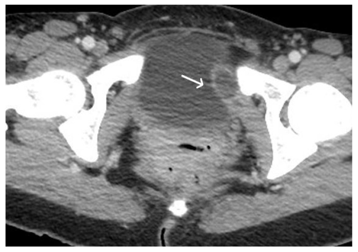
Urethral mesh placement has become a common surgical intervention for the management of stress urinary incontinence. While this procedure offers significant benefits, it is not without potential complications. This review article aims to provide a comprehensive overview of urethral mesh assessment in oncologic patients. The article explores normal magnetic resonance imaging (MRI) and computed tomography (CT) mesh appearances and highlights the pathological aspects associated with urethral mesh complications including both short-term and long-term post-operative complications. By understanding the spectrum of normal findings of urethral mesh and the possible complications, clinicians can improve patient outcomes and make informed decisions regarding urethral mesh management in this patient population.

Keywords: TOT; TVT; mesh; urethral mesh; urethral neoplasm.


Conflict of interest statement

The authors declare no conflict of interest.

Figures

Figure 1
Figure 1
TVT and TOT urethral meshes on sagittal plane.
Figure 2
Figure 2
Normal appearance of TVT mesh. Sagittal T2WI showing a thin, linear low signal intensity mesh (arrows), anchor within the subcutaneous fat, extending through the rectus sheath and inferior to the bladder within the retropubic space.
Figure 3
Figure 3
Normal appearance of TVT mesh. Axial (A) and coronal (B) T2WI demonstrating linear low signal intensity mesh anchor within the subcutaneous fat, extending through the rectus sheath (black arrows) and anteroinferior to the bladder within the retropubic space (white arrows).
Figure 4
Figure 4
Normal appearance of TOT mesh. Axial (A) and coronal (B) T2WI display a linear low signal intensity of a normal TOT mesh within the paraurethral space, posterior to the mid-urethra (U), anterior to vagina (V) and rectum (R), extending beyond the obturator foramen, towards the adductor muscles (white arrows).
Figure 5
Figure 5
Normal TVT mesh appearance on CT imaging. The mesh is seen as a thin and linear structure passing through the anterior abdominal wall (arrows).
Figure 6
Figure 6
Patient with bladder cancer and TOT mesh. Axial (A) and sagittal (B) T2WI images demonstrate extensive abnormal soft tissue of the anterior aspect of bladder (black arrows) in the setting of a normal TOT (white arrows).
Figure 7
Figure 7
Patient with ovarian cancer and TVT mesh. Sagittal (A) and axial (B) contrast enhanced CT showing a mixed cystic-solid ovarian mass (black arrow), ascites and pleural effusion. Normal TVT is visible in both planes (white arrows).
Figure 8
Figure 8
Desmoid tumour of the anterior abdominal wall, with TVT mesh. Sagittal (A) and axial (B,C) T2WI demonstrate low T2 signal desmoid tumour of the anterior abdominal wall (white arrows). Normal appearance of the TVT seen passing through the inferior aspect of the desmoid tumour (black arrows).
Figure 9
Figure 9
Acute haematoma post TVT procedure. Post-contrast CT imaging is highlighting a well-defined collection (white arrow) in left obturator internus muscle; no signs of active bleeding are noted.
Figure 10
Figure 10
Acute pelvic haematoma post sacrohysteropexy. Post-contrast CT imaging demonstrates a large, well-defined and hyperdense collection (haematoma) located within the pelvis, with important mass effect on the bladder and rectum; no signs of active bleeding are noted.
Figure 11
Figure 11
Haemoperitoneum and right rectus sheath haematoma post sacro-hysteropexy. Post-contrast CT imaging demonstrates hyperdense free fluid (*) located in the pelvic peritoneal reflection suggestive of haemoperitoneum and a well-defined hyperdense collection (white arrow) located within the right rectus abdominal muscle consistent with a haematoma; no signs of active bleeding are noted.
Figure 12
Figure 12
History of cervical cancer, TOT in situ and pubic symphysis osteomyelitis. Axial T2WI (A) and T1 fat saturated, post contrast (B) sequences demonstrate thickened left limb of TOT traversing obturator foramen to adductor muscles (black arrow), with post-contrast enhancement (white arrow) sugestive of infection. Enhancement at the pubic symphysis (white arrowheads) with loss of normal low T1 and T2 signal of the cortex (black arrowheads) consistent with osteomyelitis.
Figure 13
Figure 13
Upper images: Complicated, infected right limb of TVT and bladder erosion. Axial T2WI (A) and T1 fat saturated, post contrast (B) sequences demonstrate TVT with abscess of right limb, extending from the bladder wall to the skin surface in keeping with a sinus tract (white arrows) due to a bladder erosion. Lower images: Complicated, infected right limb of TVT. Coronal (C) and axial (D) T2WI sequences demonstrate thickened right limb of TVT within bladder wall (white arrows). Extension to skin partially visualised on axial sequence (black arrow). Artefact from right hip prosthesis.
Figure 13
Figure 13
Upper images: Complicated, infected right limb of TVT and bladder erosion. Axial T2WI (A) and T1 fat saturated, post contrast (B) sequences demonstrate TVT with abscess of right limb, extending from the bladder wall to the skin surface in keeping with a sinus tract (white arrows) due to a bladder erosion. Lower images: Complicated, infected right limb of TVT. Coronal (C) and axial (D) T2WI sequences demonstrate thickened right limb of TVT within bladder wall (white arrows). Extension to skin partially visualised on axial sequence (black arrow). Artefact from right hip prosthesis.
Figure 14
Figure 14
Infected limb of TOT and vaginal exposure. Axial T2WI (A), T1 fat saturated (B), and T2 fat saturated, post contrast (C) sequences of TOT demonstrate marked thickening and avid post contrast enhancement of infected right limb of mesh (white arrows), most likely due to a vaginal exposure.
Figure 15
Figure 15
Bladder erosion. Sagittal (A) and coronal (B) T2WI showing left arm of TVT passing through the left anterolateral bladder wall (white arrows). The bladder wall is markedly thickened at this site with irregularity and oedema of the adjacent urothelium.
Figure 16
Figure 16
Urethral erosion. Axial (A) and sagittal (B) T1 fat saturated post-contrast sequences, demonstrating linear low signal within urethra (white arrows), reflecting erosion of mesh into the urethra. There is enhancement of the lower urethra and vagina (arrowheads). Cystoscopy confirmed synthetic fibres within urethral lumen, which were resected.
Figure 17
Figure 17
Vaginal exposure. (Same patient). Axial T2WI (A) and axial fat saturated post-contrast T1 (B) images. Linear low signal intensity mesh within the oedematous vagina, compatible with mesh exposure (white arrows).
Figure 18
Figure 18
Normal clinical examination. Inflammation without mesh erosion. Axial T2WI (A) and axial fat saturated post-contrast T1WI (B) demonstrating abnormal high/intermediate T2 signal intensity of mesh as it passes between the vagina and urethra (grey arrows). On post contrast imaging, this portion of the mesh demonstrates avid enhancement (white arrows).

Similar articles

References

    1. Afonso J.S., Jorge R.M., Martins P.S., Soldi Mda S., Alves O.L., Patricio B., Mascarenhas T., Sartori M.G., Girao M.J. Structural and thermal properties of polypropylene mesh used in treatment of stress urinary incontinence. Acta Bioeng. Biomech. 2009;11:27–33. - PubMed
    1. Nolfi A.L., Brown B.N., Liang R., Palcsey S.L., Bonidie M.J., Abramowitch S.D., Moalli P.A. Host response to synthetic mesh in women with mesh complications. Am. J. Obstet. Gynecol. 2016;215:206.e1–206.e8. doi: 10.1016/j.ajog.2016.04.008. - DOI - PMC - PubMed
    1. DeLancey J.O. Structural support of the urethra as it relates to stress urinary incontinence: The hammock hypothesis. Am. J. Obstet. Gynecol. 1994;170:1713–1720; discussion 1720. doi: 10.1016/S0002-9378(12)91840-2. - DOI - PubMed
    1. Delorme E. Trans-obturator urethral suspension: A minimally invasive procedure to treat female stress urinary incontinence. Prog. Urol. 2001;11:1306–1313. - PubMed
    1. Abdel-Fattah M., Cooper D., Davidson T., Kilonzo M., Hossain M., Boyers D., Bhal K., Wardle J., N’Dow J., MacLennan G., et al. Single-Incision Mini-Slings for Stress Urinary Incontinence in Women. N. Engl. J. Med. 2022;386:1230–1243. doi: 10.1056/NEJMoa2111815. - DOI - PubMed

LinkOut - more resources