. 2023 Nov 28;15(23):5625.
doi: 10.3390/cancers15235625.

The Role of ZNF275/AKT Pathway in Carcinogenesis and Cisplatin Chemosensitivity of Cervical Cancer Using Patient-Derived Xenograft Models

Affiliations

The Role of ZNF275/AKT Pathway in Carcinogenesis and Cisplatin Chemosensitivity of Cervical Cancer Using Patient-Derived Xenograft Models

Miaomiao Ye et al. Cancers (Basel). .

Abstract

Zinc finger protein 275 (ZNF275) is a C2H2-type transcription factor that is localized on chromosome Xq28. Whether ZNF275 participates in modulating the biological behaviors of cervical cancer has not been determined to our knowledge. The present study employed CCK-8, BrdU, flow cytometry, and a transwell assay to investigate the cell viability, proliferation, apoptosis, migration, and invasion of cervical cancer cells. The application of Western blotting and immunohistochemistry (IHC) aims to assess ZNF275 protein expression and identify the signaling pathway relevant to ZNF275-mediated effects on cervical cancer. The therapeutic impact of the combined therapy of the AKT inhibitor triciribine and cisplatin was evaluated on cervical cancer patient-derived xenograft (PDX) models expressing high ZNF275. The current research illustrated that cervical cancer tissue exhibited a higher expression of ZNF275 in contrast to the surrounding normal cervical tissue. The downregulation of ZNF275 suppressed cell viability, migration, and invasion, and facilitated the apoptosis of SiHa and HeLa cells via weakening AKT/Bcl-2 signaling pathway. Moreover, triciribine synergized with cisplatin to reduce cell proliferation, migration, and invasion, and enhanced the apoptosis of SiHa cells expressing high ZNF275. In addition, the combination treatment of triciribine and cisplatin was more effective in inducing tumor regression than single agents in cervical cancer PDX models expressing high ZNF275. Collectively, the current findings demonstrated that ZNF275 serves as a sufficiently predictive indicator of the therapeutic effectiveness of the combined treatment of triciribine and cisplatin on cervical cancer. Combining triciribine with cisplatin greatly broadens the therapeutic options for cervical cancer expressing high ZNF275, but further research is needed to confirm these results.

Keywords: cervical cancer; chemosensitivity; cisplatin; patient-derived xenograft (PDX) models; zinc finger protein 275 (ZNF275).


Conflict of interest statement

The authors declare no conflict of interest.

Figures
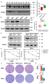
Figure 1
Figure 1
The effect of ZNF275 knockdown on viability of cervical cancer cells. (A) Left panel: ZNF275 protein expression in cervical cancer tissues and adjacent normal tissues was detected by Western blotting. N: normal tissue. T: cervical cancer tissue. Right panel: Quantitative analysis of the results. (B) The expression of ZNF275 protein in cervical cancer cell lines was determined by Western blotting. (C) The differential expression of ZNF275 in cervical cancer tissues and normal cervical tissues was retrieved in Gene Expression Omnibus (GEO) database. (D) The efficiencies of ZNF275 knockdown in SiHa and HeLa cells were validated by Western blotting. (E) CCK-8 assay revealed the effect of ZNF275 downregulation on viabilities of SiHa and HeLa cells. (F) Colony formation assay revealed the effect of ZNF275 downregulation on clonogenicity of SiHa and HeLa cells. (G) Quantitative analysis of the results in (F) of SiHa cells. (H) Quantitative analysis of the results in (F) of HeLa cells. In all panels, * p < 0.05, ** p < 0.01, *** p < 0.001, **** p < 0.0001 vs. control or normal groups. The uncropped Western blots are shown in File S1.
Figure 2
Figure 2
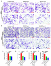
The effect of ZNF275 knockdown on migration and invasion of cervical cancer cells. (A,B) Transwell assay revealed the effect of ZNF275 downregulation on migration and invasion of SiHa (A) and HeLa (B) cells in comparison of their control groups. (C,D) Quantitative analysis of the results of migration (C) and invasion (D) in (A). (E,F) Quantitative analysis of the results of migration (E) and invasion (F) in (B). Scale bar = 100 µm. **** p < 0.0001 vs. control groups.
Figure 3
Figure 3
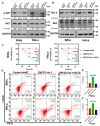
The effect of ZNF275 knockdown on apoptosis of cervical cancer cell. (A,B) Flow cytometric analysis revealed the effect of ZNF275 downregulation on apoptosis of SiHa (A) and HeLa (B) cells in comparison of their control groups. (C) Quantitative analysis of the results of (A). (D) Quantitative analysis of the results of (B). ** p < 0.01, *** p < 0.001, **** p < 0.0001 vs. control groups. (E): The relationship between ZNF275 and AKT expression in cervical cancer samples was explored in the Gene Expression Profiling Interactive Analysis (GEPIA) database. (F) The expression levels of AKT and p-AKT protein in ZNF275 knockdown SiHa and HeLa cells in comparison to their control groups were detected by Western blotting. (G) The expression levels of apoptosis-associated protein Bcl-2 and Bax in ZNF275 knockdown SiHa and HeLa cells in comparison to their control groups were detected by Western blotting. The uncropped Western blots are shown in File S1.
Figure 4
Figure 4
The effect of AKT activation by treating with 10 μM SC79 for 48 h on ZNF275 knockdown cervical cancer cells. (A) The expression levels of AKT and p-AKT protein were detected by Western blotting. (B) The expression levels of Bcl-2 and Bax protein were detected by Western blotting. (C) Left panel: The cell viability of SiHa cells was revealed by CCK-8 assay. Right panel: The cell viability of HeLa cells was revealed by CCK-8 assay. (D) The apoptosis of SiHa and HeLa cells was analyzed by flow cytometry. (E) Quantitative analysis of the results of SiHa cells in (D). (F) Quantitative analysis of the results of HeLa cells in (D). *** p < 0.001, **** p < 0.0001 vs. control groups or ZNF275 sh-1 groups. The uncropped Western blots are shown in File S1.
Figure 5
Figure 5
The effect of triciribine and cisplatin on cell viability of SiHa and Caski cells. (A) The expression of AKT and p-AKT protein in cervical cancer cell lines were determined by Western blotting. (B) Left panel: The viabilities of SiHa cells at different concentrations of triciribine (0, 50, 100, 200, and 400 μM) for 24 h, 48 h, and 72 h were determined by CCK-8 assay. Right panel: The viabilities of Caski cells at different concentrations of triciribine (0, 12.5, 25, 50, 75, and 100 μM) for 24 h, 48 h, and 72 h were determined by CCK-8 assay. (C) Left panel: The effects of triciribine (100 μM for 48 h) on AKT and p-AKT protein expression in SiHa cells were detected by Western blotting. Right panel: The effects of triciribine (12.5 μM for 48 h) on AKT and p-AKT protein expression in Caski cells were detected by Western blotting. (D) Left panel: The viabilities of SiHa cells at different concentrations of cisplatin (0, 2, 4, 6, 8, 16, and 20 μM) for 48 h were determined by CCK-8 assay. Right panel: The viabilities of Caski cells at different concentrations of cisplatin (0, 0.5, 1, 2, 4, 8, and 16 μM) for 48 h were determined by CCK-8 assay. (E) The viabilities of SiHa cells with the combined treatment of triciribine (100 μM) and different concentrations of cisplatin (0, 2, 4, and 6 μM) for 48 h were determined by CCK-8 assay. (F) The viabilities of Caski cells with the combined treatment of triciribine (12.5 μM) and different concentrations of cisplatin (0, 0.5, 1, and 2 μM) for 48 h were determined by CCK-8 assay. ** p < 0.01 vs. control group, **** p < 0.0001 vs. control group, #### p < 0.0001 vs. triciribine group. &&&& p < 0.0001 vs. cisplatin group. The uncropped Western blots are shown in File S1.
Figure 6
Figure 6
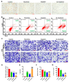
The effect of triciribine combined with cisplatin on the biological behaviors of SiHa cells. SiHa cells were treated with control (complete DMEM), triciribine (100 μM), cisplatin (4 μM), or the co-treatment of triciribine (100 μM) and cisplatin (4 μM) for 48 h. (A) The proliferation of SiHa cells was revealed by BrdU assay. Scale bar = 75 µm. (B) The apoptosis of SiHa cells was analyzed by flow cytometry (C): Transwell assay revealed the migration and invasion of SiHa cells. Scale bar = 100 µm. (D) Quantitative analysis of the results of proliferation rates in (A). (E) Quantitative analysis of the results of apoptosis in (B). (F,G) Quantitative analysis of the results of migration (F) and invasion (G) in (C). **** p < 0.0001 vs. control group, #### p < 0.0001 vs. triciribine group, &&& p < 0.001 and &&&& p < 0.0001 vs. cisplatin group.
Figure 7
Figure 7
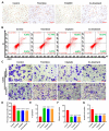
The effect of triciribine combined with cisplatin on the biological behaviors of Caski cells. Caski cells were treated with control (complete DMEM), triciribine (12.5 μM), cisplatin (1 μM), or the combination of triciribine (12.5 μM) and cisplatin (1 μM) for 48 h. (A) The proliferation of Caski cells was revealed by BrdU assay. (B) The apoptosis of Caski cells was analyzed by flow cytometry. (C) Transwell assay revealed the migration and invasion of Caski cells. (D) Quantitative analysis of the results of proliferation rates in (A). (E) Quantitative analysis of the results of apoptosis in (B). (F,G) Quantitative analysis of the results of migration (F) and invasion (G) in (C). Scale bar = 100 µm. **** p < 0.0001 vs. the control group.
Figure 8
Figure 8
The therapeutic efficacies of cisplatin, triciribine, and the co-treatment were evaluated in cervical cancer F3-PDX mice. Cervical cancer F3-PDX mice were treated with PBS (control), cisplatin (4 mg/kg), triciribine (12 mg/kg), and the co-treatment of cisplatin (4 mg/kg) and triciribine (12 mg/kg). (A) Immunohistochemistry staining revealed the protein expression of ZNF275 in patient original cervical cancer tissue (F0), paired adjacent non-cancerous cervical tissue (N0), and F2- and F3-PDX tissue (original magnification, 200×). (B) Cervical cancer xenografts were separated and photographed on the 20th day after drug treatment. (C) Tumor volumes were measured and calculated. (D) The pathology of tumor tissue was observed by hematoxylin–eosin (HE) staining (original magnification, 100×). Immunohistochemistry staining described the protein expression of ZNF275 (E), AKT (F), and p-AKT (G) in tumor tissues with different treatments (original magnification, 200×). * p < 0.05 vs. control group or triciribine group or cisplatin group.

Similar articles

Cited by

References

    1. Sung H., Ferlay J., Siegel R., Laversanne M., Soerjomataram I., Jemal A., Bray F. Global cancer statistics 2020: GLOBOCAN estimates of incidence and mortality worldwide for 36 cancers in 185 countries. CA Cancer J. Clin. 2021;71:209–249. doi: 10.3322/caac.21660. - DOI - PubMed
    1. Small W., Jr., Bacon M.A., Bajaj A., Chuang L.T., Fisher B.J., Harkenrider M.M., Jhingran A., Kitchener H.C., Mileshkin L.R., Viswanathan A.N., et al. Cervical cancer: A global health crisis. Cancer. 2017;123:2404–2412. doi: 10.1002/cncr.30667. - DOI - PubMed
    1. Masadah R., Rauf S., Pratama M.Y., Tiribelli C., Pascut D. The Role of microRNAs in the Cisplatin- and Radio-Resistance of Cervical Cancer. Cancers. 2021;13:1168. doi: 10.3390/cancers13051168. - DOI - PMC - PubMed
    1. Cohen A., Roane B., Leath C. Novel Therapeutics for Recurrent Cervical Cancer: Moving Towards Personalized Therapy. Drugs. 2020;80:217–227. doi: 10.1007/s40265-019-01249-z. - DOI - PMC - PubMed
    1. Tian X., Wang X., Cui Z., Liu J., Huang X., Shi C., Zhang M., Liu T., Du X., Li R., et al. A Fifteen-Gene Classifier to Predict Neoadjuvant Chemotherapy Responses in Patients with Stage IB to IIB Squamous Cervical Cancer. Adv. Sci. 2021;8:2001978. doi: 10.1002/advs.202001978. - DOI - PMC - PubMed

LinkOut - more resources