. 2023 Nov 29;15(23):5651.
doi: 10.3390/cancers15235651.

HSP110 Inhibition in Primary Effusion Lymphoma Cells: One Molecule, Many Pro-Survival Targets

Affiliations

HSP110 Inhibition in Primary Effusion Lymphoma Cells: One Molecule, Many Pro-Survival Targets

Roberta Gonnella et al. Cancers (Basel). .

Abstract

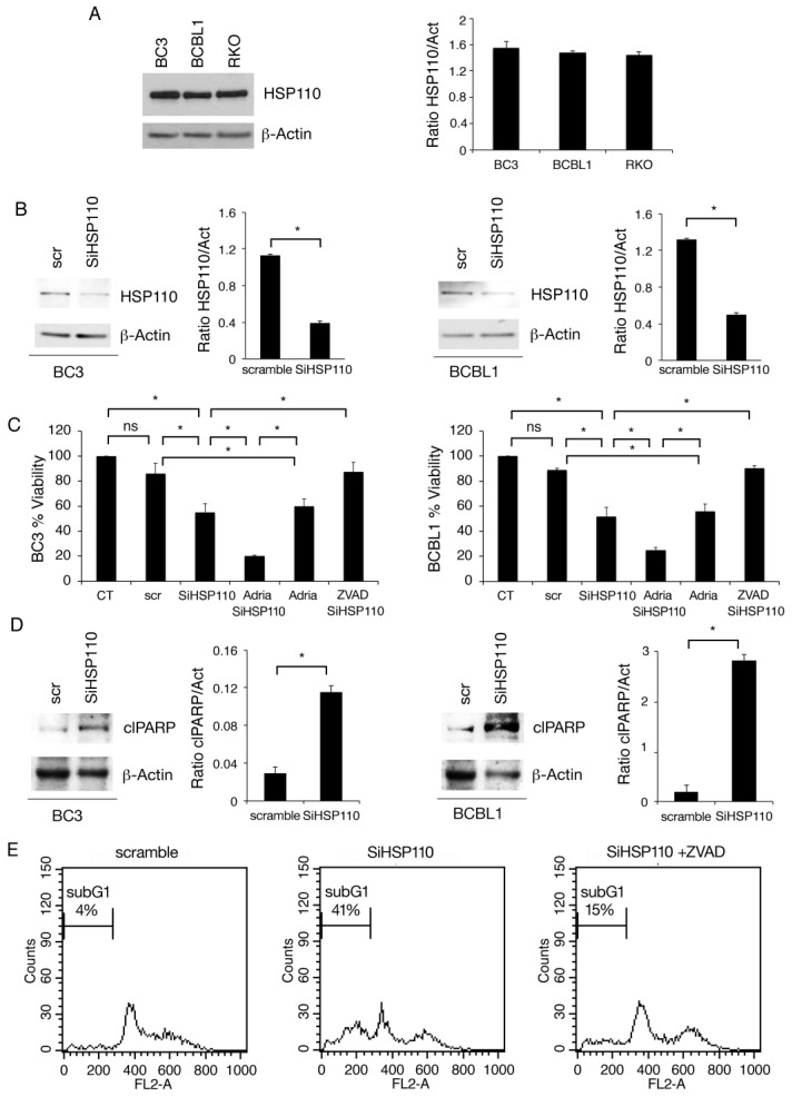
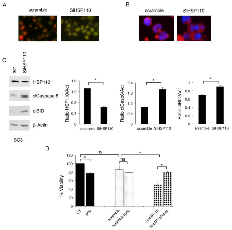
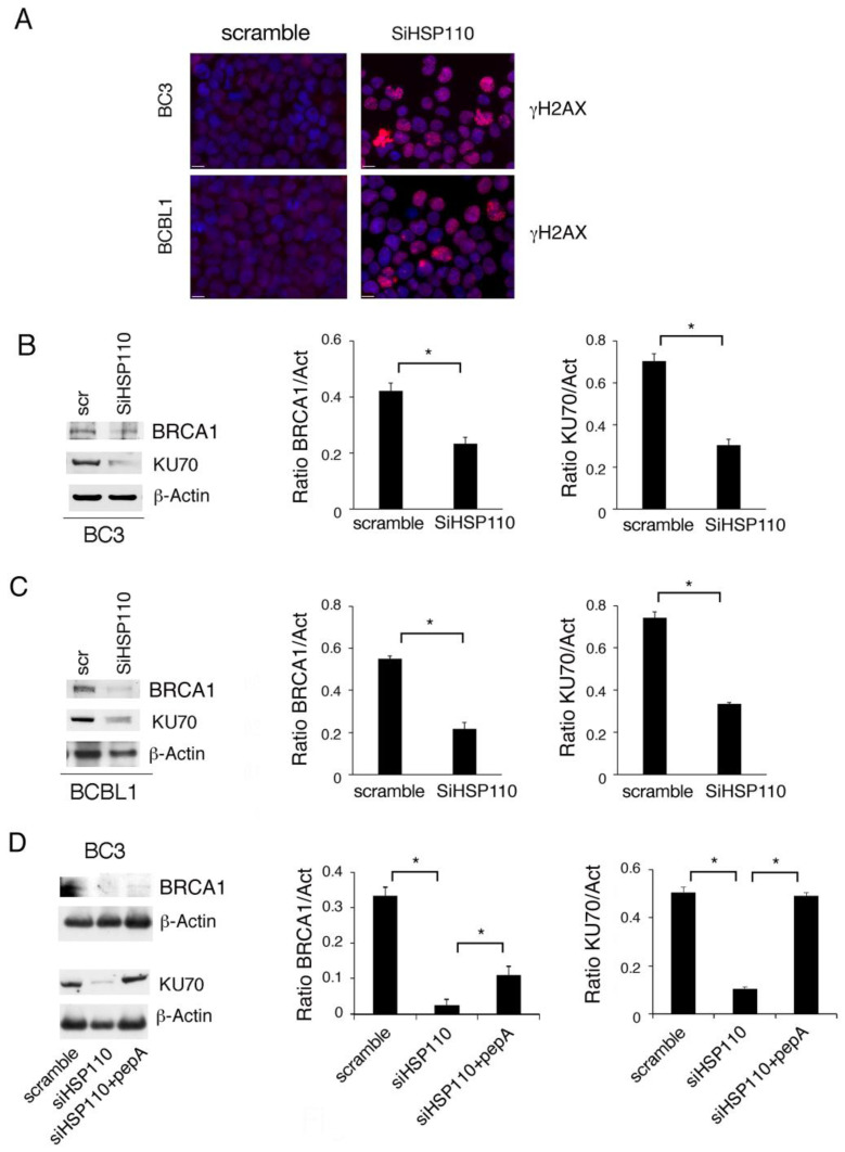
Heat shock proteins (HSPs) are highly expressed in cancer cells and represent a promising target in anti-cancer therapy. In this study, we investigated for the first time the expression of high-molecular-weight HSP110, belonging to the HSP70 family of proteins, in Primary Effusion Lymphoma (PEL) and explored its role in their survival. This is a rare lymphoma associated with KSHV, for which an effective therapy remains to be discovered. The results obtained from this study suggest that targeting HSP110 could be a very promising strategy against PEL, as its silencing induced lysosomal membrane permeabilization, the cleavage of BID, caspase 8 activation, downregulated c-Myc, and strongly impaired the HR and NHEJ DNA repair pathways, leading to apoptotic cell death. Since chemical inhibitors of this HSP are not commercially available yet, this study encourages a more intense search in this direction in order to discover a new potential treatment that is effective against this and likely other B cell lymphomas that are known to overexpress HSP110.

Keywords: DDR; HSP110; PEL; apoptosis.


Conflict of interest statement

The authors declare no conflict of interest.

Figures

Figure 1
Figure 1
HSP110 is highly expressed in PEL cells and sustains cell survival. (A) Protein expression level of HSP110 was evaluated via Western blot analysis in BC3, BCBL1 (PEL), and RKO (colon cancer) cell lines. PEL cells were knocked down for HSP110 or scramble-treated (scr) for 48 h; (B) the expression of HSP110 was evaluated via Western blot analysis; (C) cell survival was assessed via Trypan blue assay in PEL cells silenced for HSP110 treated with Adriamycin (Adria) or the pan-caspase inhibitor, z-VAD fmk (Z-VAD). The histograms represent the percentage of cell viability relative to the control; data are represented as the mean plus SD of more than three experiments. * p value < 0.05, as calculated using an ANOVA test. (D) Protein expression level of cleaved PARP (clPARP) was evaluated via Western blot analysis in PEL cells knocked down for HSP110 or scramble-treated (scr). (A,B,D) β-Actin was used as loading control. Histograms represent the mean plus SD of the densitometric analysis of the ratio of HSP110/β-Actin and cleaved-PARP/β-Actin (clPARP/Act). (E) Sub-G1 events as evaluated via FACS analysis in BC3 cells scramble-treated or silenced for HSP110, in the presence or in the absence of the pan-caspase inhibitor, z-VAD fmk (Z-VAD). A representative experiment is shown, and the means plus standard deviations of the densitometric analysis of three independent experiments are also reported. * p value < 0.05. The uncropped blots are shown in Supplementary Materials.
Figure 2
Figure 2
Lysosomal permeabilization contributes to the cytotoxic effect of HSP110 inhibition. (A) Lysosomal permeabilization following gene silencing of HSP110 in BC3 cells was assessed via acridine orange (AO) staining. In scramble-treated cells, AO stains lysosomes red since they have intact membranes that maintain an acidic compartment within them. In cells knocked down for HSP110 (SiHSP110), the staining becomes green due to the increase in the pH from lysosomal membrane damage. (B) Cellular localization of Cathepsin D in BC3 cells following silencing of HSP110 (SiHSP110) or scramble treatment (scramble) was evaluated in terms of immunofluorescence. (C) Western blot analysis of protein expression level of HSP110, cleaved Caspase8 (clCaspase 8), and cleaved BID (clBID) in PEL cells knocked down for HSP110 and scramble-treated (scr) for 48 h. β-Actin was used as loading control. Histograms represent the mean plus SD of the densitometric analysis of the ratio of HSP110/β-Actin, cleaved-Caspase8/β-Actin (clCaspase8/Act), and cleaved BID/β-Actin (clBID/Act). A representative experiment is shown, and the means plus standard deviations of the densitometric analysis of three independent experiments are also reported. * p value < 0.05, as calculated via ANOVA test. (D) Cell survival of BC3 cells following HSP110 gene silencing, in combination or not with the cathepsin inhibitor, Pepstatin A (pep), assessed using Trypan blue assay. The histograms represent the percentage of cell viability relative to the control; data are represented as the mean plus SD of more than three experiments. * p value < 0.05.
Figure 3
Figure 3
HSP110 knockdown induces DNA damage by impairing DDR molecules. (A) The effect of HSP110 gene silencing (SiHSP110) on DNA damage in BC3 and BCBL1 cells was evaluated in terms of immunofluorescence of γH2AX foci. Bars = 10 μm. (B,C) Western blot analysis of BRCA1 and KU70 protein expression in PEL cells knocked down for HSP110 and scramble-treated (scr) for 48 h. β-Actin was used as loading control. Histograms represent the mean plus SD of the densitometric analysis of the ratio of HSP110/β-Actin (HSP110/Act), BRCA1/β-Actin (BRCA1/Act), and KU70/β-Actin (KU70/Act). (D) Protein expression level of BRCA1 and KU70 was evaluated in BC3 cells knocked down for HSP110 untreated or treated with Pepstatin A. β-Actin was used as loading control. Histograms represent the mean plus SD of the densitometric analysis of the ratio of BRCA1/β-Actin (BRCA1/Act) and KU70/β-Actin (KU70/Act). (BD) A representative experiment is shown, and the means plus standard deviations of the densitometric analysis of three independent experiments are also reported. * p value < 0.05.
Figure 4
Figure 4
HSP110 silencing induces c-Myc and phospho-STAT3 downregulation, p21 upregulation, and DNA damage induction in PEL cells. (A,B) Western blot analysis of c-Myc, phospho-STAT3 (Tyr705), and STAT3 protein expression in PEL cells knocked down for HSP110 (SiHSP110) or scramble-treated (scr) for 48 h. (C) Western blot analysis of p21 protein expression in BC3 cells following HSP110 gene silencing. (D) BRCA1 and KU70 protein expression level was evaluated via Western blot analysis in BC3 cells treated with c-Myc inhibitor (I c-Myc). β-Actin was used as loading control. Histograms represent the mean plus SD of the densitometric analysis of the ratio of HSP110/β-Actin (HSP110/Act), c-Myc/β-Actin (c-Myc/Act), phospho-STAT3/β-Actin (pSTAT3/Act), STAT3/β-Actin (STAT3/Act), p21/β-Actin, BRCA1/β−Actin (BRCA1/Act), and KU70/β-Actin (KU70/Act). A representative experiment is shown, and the means plus standard deviations of the densitometric analysis of three independent experiments are also reported. * p value < 0.05.

Similar articles

Cited by

References

    1. Yang S., Xiao H., Cao L. Recent advances in heat shock proteins in cancer diagnosis, prognosis, metabolism and treatment. Biomed. Pharmacother. 2021;142:112074. doi: 10.1016/j.biopha.2021.112074. - DOI - PubMed
    1. Chen M., Xie S. Therapeutic targeting of cellular stress responses in cancer. Thorac. Cancer. 2018;9:1575–1582. doi: 10.1111/1759-7714.12890. - DOI - PMC - PubMed
    1. Briones J. Heat-shock proteins: A c-Myc lymphoma target? Blood. 2015;125:1685–1686. doi: 10.1182/blood-2015-01-622191. - DOI - PubMed
    1. Alexandrova E.M., Marchenko N.D. Mutant p53—Heat Shock Response Oncogenic Cooperation: A New Mechanism of Cancer Cell Survival. Front. Endocrinol. 2015;6:53. doi: 10.3389/fendo.2015.00053. - DOI - PMC - PubMed
    1. Gilardini Montani M.S., Cecere N., Granato M., Romeo M.A., Falcinelli L., Ciciarelli U., D’Orazi G., Faggioni A., Cirone M. Mutant p53, Stabilized by Its Interplay with HSP90, Activates a Positive Feed-Back Loop Between NRF2 and p62 that Induces Chemo-Resistance to Apigenin in Pancreatic Cancer Cells. Cancers. 2019;11:703. doi: 10.3390/cancers11050703. - DOI - PMC - PubMed

LinkOut - more resources