Review
. 2023 Dec 2;15(23):5694.
doi: 10.3390/cancers15235694.

Unveiling the Immunogenicity of Ovarian Tumors as the Crucial Catalyst for Therapeutic Success

Affiliations
Review

Unveiling the Immunogenicity of Ovarian Tumors as the Crucial Catalyst for Therapeutic Success

Galaxia M Rodriguez et al. Cancers (Basel). .

Abstract

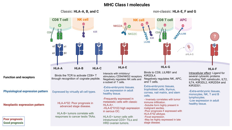
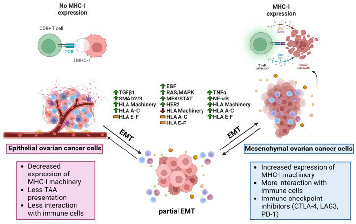
Epithelial ovarian cancer (EOC) is the most lethal gynecologic cancer. The disease is often diagnosed after wide-spread dissemination, and the standard treatment combines aggressive surgery with platinum-based chemotherapy; however, most patients experience relapse in the form of peritoneal carcinomatosis, resulting in a 5-year mortality below 45%. There is clearly a need for the development of novel treatments and cancer immunotherapies offering a different approach. Immunotherapies have demonstrated their efficacy in many types of cancers; however, only <15% of EOC patients show any evidence of response. One of the main barriers behind the poor therapeutic outcome is the reduced expression of Major Histocompatibility Complexes class I (MHC I) which occurs in approximately 60% of EOC cases. This review aims to gather and enhance our current understanding of EOC, focusing on its distinct cancer characteristics related to MHC I expression, immunogenicity, antigen presentation, epithelial-to-mesenchymal transition, and various ongoing immunotherapeutic strategies designed to stimulate antitumor immunity.

Keywords: EMT; classic HLA I; non-classic HLA I; ovarian cancer; tumor immunogenicity; tumor-associated antigens.


Conflict of interest statement

The authors declare no conflict of interest.

Figures

Figure 1
Figure 1
Classic and non-classic HLA I and their role in EOC tumorigenicity. HLA I molecules are formed from three α subunits (α1-3) and one B2M subunit (β2). Classic HLA I molecules include HLA-A, -B and –C while non-classic are known as HLA-E, -F, G. Classic HLA I molecules allow the presentation of endogenous peptides to CD8+ T cells, while non-classic HLA I molecules negatively regulate NK cell function. Some classic HLA Is (HLA-A*02) have been associated with poor EOC prognosis (red captions) when co-expressed with non- classic HLA-G and HLA-E [8]. HLA-E can interact with stimulatory or inhibitory receptors on NK cells and some T cells, while HLA-Gs have a broader immune-modulatory capability, having a negative effect on NKs, T cells and APCs. sHLA-G can also be found in extracellular vesicles and tumor derived exosomes. HLA-E and HLA-G expression levels have been associated with poor prognosis and late stage of disease. However, in some specific cases, HLA-E expression along T cell infiltration and HRD ovarian tumors have shown a good prognosis (green captions). HLA-F may have immune regulatory functions, but its binding partners are still unknown. See text for more details. HLA-A*02 refers to the allele group, TCR (T cell receptor), NK (natural killer cell), antigen presenting cell (APC), OC (ovarian cancer), HRD (homologous recombinant deficiency), TAP (transporter associated with antigen presentation), ILT2 [immunoglobulin (Ig)-like transcript 2], KIR3DS1 (killer cell Ig like receptor three Ig domains and short cytoplasmic tail 1), KIR3DL2 (KIR3D and long cytoplasmic tail 2), KIR2DS4 (KIR two Ig domains and short cytoplasmic tail 4), LILRB1 (Leukocyte Immunoglobulin-Like Receptor B1).
Figure 2
Figure 2
Average gene expression of MHC-I related HLA allotypes correlates with EMT in various cancer types. Expression of MHC-I related HLA allotypes (HLA A-G) scored in the cancer cell compartment of a variety of tumors (BC—Breast Cancer, CC—Colon Cancer, GC—Gastric Cancer, LC—Lung Cancer, NPC—Nasopharyngeal Cancer, OC—Ovarian Cancer, PDAC—Pancreatic Ductal Adenocarcinoma, SCC—Squamous Cell Carcinoma). HLA expression was correlated with EMT scores of those cells based on either (A) a cancer-specific EMT module published by Cook and Vanderhyden [120] or (B) the Hallmark EMT module from MSigDb.
Figure 3
Figure 3
Average gene expression of HLA I allotypes and some APM components correlate with EMT in cancer cell lines. Heat map showing expression levels of transcripts for MHC-I related HLA allotypes (HLA A-G) and machinery (TAP1, TAP2, PSMB9) in ovarian (OVCA420), breast (MCF7), and lung (A549) cancer cell lines in response to treatment with (A) TGF β1, (B) EGF, (C) TNFα for 0 and 8 h, and 1, 3, and 7 days. Data were extracted from single-cell RNA-seq datasets published by Cook and Vanderhyden [130].
Figure 4
Figure 4
Therapeutic interventions aiming to increase EOC immunogenicity. TAAs highly expressed by EOC cells such as HER2, MSLN, CA125, FRα, and TAG42/CD47 can be targeted with CAR-T cells. FRα can be targeted by direct binding with antibody-drug conjugates. HLA-E+ ovarian tumors can be treated with antibodies blocking the HLA-E/NKG2A axis and impeding NK cell anti-tumoral activity. Poorly immunogenic ovarian tumors can be treated with DNMTi or HDACi agents aiming to decrease DNA hypermethylation or histone deacetylation (indicated by the red “X”). These epigenetic modifiers promote an increase in MHC I gene expression, type I IFN release, and TAA presentation, increasing CTL binding and antitumoral activity. HLA-G+ ovarian tumors can be treated with CAR-NK cells aiming to restore NK cytolytic functions. Green (+) symbols denote activation effect while red (−) symbols denote inhibitory signaling. NK (Natural Killer cell), CAR (Chimeric Antigen Receptor), HER2 (Human Epidermal Growth Factor Receptor 2), MSLN (Mesothelin), CA125 (Cancer Antigen 125), FRα (Alpha-Folate Receptor), TAG72 (Tumor-associated glycoprotein 72), NKG2A (NK group 2 member A receptor), CTLs (Cytotoxic T Lymphocytes), DNMTi (DNA methyltransferase inhibitor), HDACi (histone deacetylase inhibitor), IRF-1 (Interferon Regulatory Factor 1), ISRE (Interferon-Stimulated Response Element), NLRC5 (NLR family CARD domain containing 5).
Figure 5
Figure 5
Potential relationship between antigen presentation and EMT in ovarian cancer. In ovarian cancer, epithelial cancer cells have decreased expression of classical HLA I related machinery, less interaction with immune cells, and less TAA presentation. Once stimulated with TGF-β1, EMT can begin increasing the expression of EMT-TFs such as SNAIL, SLUG, ZEB1, and TWIST1/2. At the same time, SMAD2/3 pathways are active, leading to the activation of APM and the re-expression of HLA-A, -B, and -C, and further leading to increased immunogenicity as the cells undergo EMT. If the EGF receptor (EGFR) is stimulated, the cells begin to undergo an EMT coinciding with activation of the RAS/MAPK, MEK/STAT, and HER2 pathways while seeing a reduction in APM and no changes to typical and atypical HLA I allotypes. EMT also beings if cancer cells are stimulated by TNF-α, including an increase in NF-κB that is upstream of APM and both typical and atypical HLA molecules. With increased immunogenicity in the mesenchymal phenotype as a result of more antigen presentation, immune cells like CD8+ T cells are able to effectuate their cytolytic functions on the cancer cells through PFN, GZMB, IFN-γ, and TNF-α. TAA (Tumor associated antigen), TGF-β1 (Transforming growth factor β1), EMT-TF (Epithelial-mesenchymal transition transcription factor), SNAIL (Zinc finger protein SNAI1), SLUG (Zinc finger protein SNAI2), ZEB1 (Zinc finger E-box binding homeobox1), TWIST1/2 (Twist-related protein 1/2), SMAD2/3 (Mothers against decapentaplegic homolog 2/3), APM (Antigen presenting machinery), HLA (Human leukocyte antigen), EGF (Epidermal growth factor), RAS/MAPK (Reticular activating system/Mitogen-activated protein kinase), MEK/STAT (Mitogen-activated protein kinase kinase/Signal transducer and activator of transcription), HER2 (Receptor tyrosine-protein kinase erbB-2), EMT (Epithelial-mesenchymal transition), NF-κB (Nuclear factor kappa-light-chain-enhancer of activated B cells), PFN (Perforin), GZMB (Granzyme B), IFN-γ (Interferon gamma).

Similar articles

Cited by

References

    1. Shiina T., Hosomichi K., Inoko H., Kulski J.K. The HLA Genomic Loci Map: Expression, Interaction, Diversity and Disease. J. Hum. Genet. 2009;54:15–39. doi: 10.1038/jhg.2008.5. - DOI - PubMed
    1. Garrido F. HLA Class-I Expression and Cancer Immunotherapy. Adv. Exp. Med. Biol. 2019;1151:79–90. doi: 10.1007/978-3-030-17864-2_3. - DOI - PubMed
    1. Klein J., Sato A. The HLA System. First of Two Parts. N. Engl. J. Med. 2000;343:702–709. doi: 10.1056/NEJM200009073431006. - DOI - PubMed
    1. Klein J., Sato A. The HLA System. Second of Two Parts. N. Engl. J. Med. 2000;343:782–786. doi: 10.1056/NEJM200009143431106. - DOI - PubMed
    1. Cruz-Tapias P., Castiblanco J., Anaya J.-M. Autoimmunity: From Bench to Bedside [Internet] El Rosario University Press; Bogotá, Colombia: 2013. HLA Association with Autoimmune Diseases. - PubMed