. 2023 Nov 21;28(23):7697.
doi: 10.3390/molecules28237697.

Design and Synthesis of Novel α-Methylchalcone Derivatives, Anti-Cervical Cancer Activity, and Reversal of Drug Resistance in HeLa/DDP Cells

Affiliations

Design and Synthesis of Novel α-Methylchalcone Derivatives, Anti-Cervical Cancer Activity, and Reversal of Drug Resistance in HeLa/DDP Cells

Zheng Yang et al. Molecules. .

Abstract

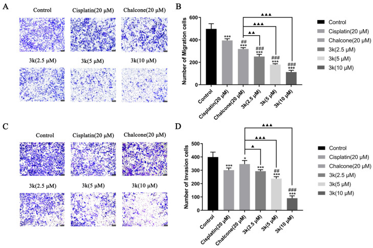
In this study, a collection of newly developed α-methylchalcone derivatives were synthesized and assessed for their inhibitory potential against human cervical cancer cell lines (HeLa, SiHa, and C33A) as well as normal human cervical epithelial cells (H8). Notably, compound 3k exhibited substantial inhibitory effects on both HeLa and HeLa/DDP cells while demonstrating lower toxicity toward H8 cells. Furthermore, the compound 3k was found to induce apoptosis in both HeLa and HeLa/DDP cells while also inhibiting the G2/M phase, resulting in a decrease in the invasion and migration capabilities of these cells. When administered alongside cisplatin, 3k demonstrated a significant reduction in the resistance of HeLa/DDP cells to cisplatin, as evidenced by a decrease in the resistance index (RI) value from 7.90 to 2.10. Initial investigations into the underlying mechanism revealed that 3k did not impact the expression of P-gp but instead facilitated the accumulation of rhodamine 123 in HeLa/DDP cells. The results obtained from CADD docking analysis demonstrated that 3k exhibits stable binding to microtubule proteins and P-gp targets, forming hydrogen bonding interaction forces. Immunofluorescence analysis further revealed that 3k effectively decreased the fluorescence intensity of α and β microtubules in HeLa and HeLa/DDP cells, resulting in disruptions in cell morphology, reduction in cell numbers, nucleus coagulation, and cell rupture. Additionally, Western blot analysis indicated that 3k significantly reduced the levels of polymerized α and β microtubule proteins in both HeLa and HeLa/DDP cell lines while concurrently increasing the expression of dissociated α and β microtubule proteins. The aforementioned findings indicate a potential correlation between the inhibitory effects of 3k on HeLa and HeLa/DDP cells and its ability to inhibit tubulin and P-gp.

Keywords: P-gp; cervical cancer; cisplatin resistance; tubulin; α-methyl chalcone.


Conflict of interest statement

The authors declare no conflict of interest.

Figures

Figure 1
Figure 1
The chemical structures of chalcone and α-methyl chalcone derivatives (14) reported in the above literature are schematically illustrated; (Chalcone: (E)-chalcone; 1: (E)-3-(6-methoxy-3a,7a-dihydro-1H-indol-3-yl)-2-methyl-1-(3,4,5-trimethoxyphenyl)prop-2-en-1-one; 2: sodium (E)-(6-methoxy-3-(2-methyl-3-oxo-3-(3,4,5-trimethoxyphenyl)prop-1-en-1-yl)-3a,7a-dihydro-1H-indol-1-yl)phosphonate; 3: (E)-1-(2,5-dimethoxyphenyl)-3-(4-(dimethylamino)phenyl)-2-methylprop-2-en-1-one; 4: (E)-3-(3a,7a-dihydro-1H-indol-3-yl)-2-methyl-1-(3,4,5-trimethoxyphenyl)prop-2-en-1-one). Red boxes represent methoxy substitution.
Figure 2
Figure 2
Schematic diagram of the rationale for the present study.
Figure 2
Figure 2
Schematic diagram of the rationale for the present study.
Scheme 1
Scheme 1
The synthetic route to target compounds 3a3p.
Scheme 2
Scheme 2
IC50 values of the lead compound chalcone, the positive drug cisplatin, and compound 3k on cervical cancer cell lines. All the above results were expressed as the mean ± SD of three independent experiments: * p < 0.05, ** p < 0.01, *** p < 0.001 compared with the chalcone group; # p < 0.05, ## p < 0.01, ### p < 0.001 compared with the cisplatin group.
Scheme 3
Scheme 3
(A) Pro-apoptotic effect of compound 3k on HeLa cells after 24 h of action. (B) Quantitative analysis of the pro-apoptotic effect. (C) Changes in the expression of pro-apoptotic protein Bax and anti-apoptotic protein Bcl-2 after 24 h of compound 3k action on HeLa cells. (D) Quantitative analysis graph of the expression of pro-apoptotic protein Bax and anti-apoptotic protein Bcl-2. All the above results were expressed as the mean ± SD of three independent experiments: * p < 0.05, ** p < 0.01, *** p < 0.001 compared with the control group; ### p < 0.001 compared with the cisplatin group; ▲ p < 0.05, ▲▲ p < 0.01, ▲▲▲ p < 0.001 compared with the chalcone group.
Scheme 4
Scheme 4
(A) Cell cycle effects of 3k and cisplatin on HeLa. (B) Quantitative analysis of the effect of 3k on the HeLa cell cycle in the bar graph. All the above results were expressed as the mean ± SD of three independent experiments: * p < 0.05, ** p < 0.01, *** p < 0.001 compared with the control group; # p < 0.05, ## p < 0.01, ### p < 0.001 compared with the cisplatin group; ▲ p < 0.05, ▲▲ p < 0.01, ▲▲▲ p < 0.001 compared with the chalcone group.
Scheme 5
Scheme 5
(A) Morphology of migrating cells after 24 h of compound 3k on HeLa cells in a Transwell migration assay. (B) Quantitative analysis of the number of migrated cells. (C) Morphology of invaded cells after 24 h of compound 3k action on HeLa cells in the Transwell invasion assay. (D) Quantitative analysis of the number of invaded cells; HeLa cell morphology was observed under an inverted microscope (magnification: ×100). All the above results were expressed as the mean ± SD of three independent experiments: * p < 0.05, *** p < 0.001 compared with the control group; # p < 0.05, compared with the cisplatin group; ▲ p < 0.05, ▲▲ p < 0.01, ▲▲▲ p < 0.001 compared with the chalcone group.
Scheme 6
Scheme 6
IC50 values of the lead compound chalcone, the positive drug cisplatin and compound 3k on cervical cancer cell lines. All the above results were expressed as the mean ± SD of three independent experiments: ** p < 0.01, *** p < 0.001 compared with the HeLa/DDP group.
Scheme 7
Scheme 7
(A) P-gp protein expression after applying Compound 3k to HeLa/DDP cells for 48 h. (B) Quantitative analysis of P-gp protein expression in HeLa/DDP cells. (C) Fluorescence intensity of rhodamine 123 after applying Compound 3k to HeLa/DDP cells for 48 h. (D) Quantitative analysis of fluorescence intensity of rhodamine 123 in HeLa/DDP cells. All the above results were expressed as the mean ± SD of three independent experiments: ** p < 0.01, *** p < 0.001 compared with the control group; ## p < 0.01 compared with the verapamil group.
Scheme 8
Scheme 8
(A) Pro-apoptotic effect of compound 3k on HeLa/DDP cells after 24 h of action. (B) Quantification of the pro-apoptotic effect on HeLa/DDP cells. (C) Changes in the expression of pro-apoptotic protein Bax and anti-apoptotic protein Bcl-2 in HeLa cells after 24 h of compound 3k action. (D) Quantification of the expression of pro-apoptotic protein Bax and anti-apoptotic protein Bcl-2 in HeLa/DDP cells. All the above results were expressed as the mean ± SD of three independent experiments: * p < 0.05, ** p < 0.01, *** p < 0.001 compared with the control group; ### p < 0.001 compared with the cisplatin group; ▲ p < 0.05, ▲▲ p < 0.01, ▲▲▲ p < 0.001 compared with the chalcone group.
Scheme 9
Scheme 9
(A) Cell cycle effects of 3k and cisplatin on HeLa/DDP. (B) Quantitative analysis of the effect of 3k on the HeLa cell cycle in the bar graph. All the above results were expressed as the mean ± SD of three independent experiments: * p < 0.05, ** p < 0.01, *** p < 0.001 compared with the control group; ▲ p < 0.05, ▲▲▲ p < 0.001 compared with the chalcone group.
Scheme 10
Scheme 10
(A) Morphology of migrated cells 24 h after compound 3k was applied to HeLa/DDP cells in the Transwell migration assay. (B) Quantitative analysis of the number of migrated cells. (C) Morphology of invaded cells 24 h after compound 3k was applied to HeLa/DDP cells in the Transwell invasion assay. (D) Quantitative analysis of the number of invaded cells; HeLa cell morphology was observed under an inverted microscope (magnification: ×100). All the above results were expressed as the mean ± SD of three independent experiments: * p < 0.05, *** p < 0.001 compared with the control group; ## p < 0.01, ### p < 0.001 compared with the cisplatin group; ▲ p < 0.05, ▲▲ p < 0.01, ▲▲▲ p < 0.001 compared with the chalcone group.
Scheme 11
Scheme 11
(A1,A2,B1,B2) 3D schematic of molecular docking of the pilot compound chalcone with tubulin and P-gp proteins; (C1,C2,D1,D2) 3D schematic of compound 3k docked with tubulin and P-gp protein molecules.
Scheme 12
Scheme 12
Effect of compound 3k on microtubulin in HeLa cells (blue fluorescence for nucleus, green fluorescence for α-tubulin, red fluorescence for β-tubulin; ×400).
Scheme 13
Scheme 13
(A) Expression level of α-/β-tubulins in polymerized state in HeLa cells. (B) Histogram of microtubulin expression level (n = 3). (C) Expression level of α-/β-tubulins in dissociated state in HeLa cells. (D) Histogram of microtubulin expression level (n = 3). All the above results were expressed as the mean ± SD of three independent experiments: * p < 0.05, ** p < 0.01, compared with the control group; # p < 0.05, ## p < 0.01, compared with the colchicine group.
Scheme 14
Scheme 14
Effect of compound 3k on microtubulin in HeLa/DDP cells (blue fluorescence for nucleus, green fluorescence for α-tubulin, red fluorescence for β-tubulin; ×400).
Scheme 15
Scheme 15
(A) Expression level of α-/β-tubulins in polymerized state in HeLa/DDP cells. (B) Histogram of microtubulin expression level (n = 3). (C) Expression level of α-/β-tubulins in dissociated state in HeLa/DDP cells. (D) Histogram of microtubulin expression level (n = 3). All the above results were expressed as the mean ± SD of three independent experiments: * p < 0.05, ** p < 0.01, *** p < 0.001 compared with the control group; ## p < 0.01 compared with the colchicine group.

Similar articles

Cited by

References

    1. Sung H., Ferlay J., Siegel R.L., Laversanne M., Soerjomataram I., Jemal A., Bray F. Global cancer statistics 2020: Globocan estimates of incidence and mortality worldwide for 36 cancers in 185 countries. CA Cancer J. Clin. 2021;71:209–249. doi: 10.3322/caac.21660. - DOI - PubMed
    1. Liu X., Xing Y., Li M., Zhang Z., Wang J., Ri M., Jin C., Xu G., Piao L., Jin H., et al. Licochalcone a inhibits proliferation and promotes apoptosis of colon cancer cell by targeting programmed cell death-ligand 1 via the nf-κb and ras/raf/mek pathways. J. Ethnopharmacol. 2021;273:113989. doi: 10.1016/j.jep.2021.113989. - DOI - PubMed
    1. Li X., Li M., Huang M., Lin Q., Fang Q., Liu J., Chen X., Liu L., Zhan X., Shan H., et al. The multi-molecular mechanisms of tumor-targeted drug resistance in precision medicine. Biomed. Pharmacother. 2022;150:113064. doi: 10.1016/j.biopha.2022.113064. - DOI - PubMed
    1. Gottesman M.M., Fojo T., Bates S.E. Multidrug resistance in cancer: Role of atp-dependent transporters. Nat. Rev. Cancer. 2002;2:48–58. doi: 10.1038/nrc706. - DOI - PubMed
    1. Doherty B., Lawlor D., Gillet J.P., Gottesman M., O’Leary J.J., Stordal B. Collateral sensitivity to cisplatin in kb-8-5-11 drug-resistant cancer cells. Anticancer Res. 2014;34:503–507. - PubMed