. 2023 Nov 24;28(23):7747.
doi: 10.3390/molecules28237747.

Computational Modeling Study of the Binding of Aging and Non-Aging Inhibitors with Neuropathy Target Esterase

Affiliations

Computational Modeling Study of the Binding of Aging and Non-Aging Inhibitors with Neuropathy Target Esterase

Wenxiong Wu et al. Molecules. .

Abstract

Neuropathy target esterase (NTE) is a serine hydrolase with phospholipase B activity, which is involved in maintaining the homeostasis of phospholipids. It can be inhibited by aging inhibitors such as some organophosphorus (OP) compounds, which leads to delayed neurotoxicity with distal degeneration of axons. However, the detailed binding conformation of aging and non-aging inhibitors with NTE is not known. In this study, new computational models were constructed by using MODELLER 10.3 and AlphaFold2 to further investigate the inhibition mechanism of aging and non-aging compounds using molecular docking. The results show that the non-aging compounds bind the hydrophobic pocket much deeper than aging compounds and form the hydrophobic interaction with Phe1066. Therefore, the unique binding conformation of non-aging compounds may prevent the aging reaction. These important differences of the binding conformations of aging and non-aging inhibitors with NTE may help explain their different inhibition mechanism and the protection of non-aging NTE inhibitors against delayed neuropathy.

Keywords: computational modeling; molecular docking; neuropathy target esterase.


Conflict of interest statement

The authors declare no conflict of interest.

Figures

Figure 1
Figure 1
The constructed NEST models. (A) The sequence alignment results between PlpD and NEST. Identical residues have been boxed in the black color. Secondary structures of PlpD (shown on the top of the sequence) and two NEST models built by MODELLER 10.3 and the AlphaFold2 (shown below the sequence) are displayed. β-strands are shown as arrows colored in cream and α-helices are shown as rectangles colored in blue. (B,C) The overall structure models of NEST built by MODELLER 10.3 (B) and AlphaFold2 (C). (D,E) The overlay of AlphaFold model and MODELLER model (D) and the overlay of the MODELLER model with the PlpD structure (E). The PlpD structure, the MODELLER model, and the AlphaFold model are colored blue, green, and cream, respectively.
Figure 2
Figure 2
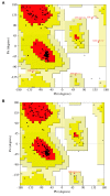
The assessment of the models of NEST. (A) Ramachandran Plot and Procheck result of the homology model built by MODELLER 10.3, in which 98.6% of the amino acid residues are in the reasonable zone. (B) Ramachandran Plot and Procheck result of the model built by the AlphaFold2, in which 100% of the amino acid residues are in the reasonable zone. In this Ramachandran Plot, the “most favored” regions are colored in red, “additional allowed” regions are colored in deep yellow, and the “generously allowed” regions are colored in light yellow while “disallowed” regions are colored in white. One black point represents one residue. Triangles indicate the glycine residues while rectangles indicate other residues. “A”, “a” and “~a” indicate the “most favored”, “additional allowed” and “generously allowed” regions of right-handed alpha helices, respectively. “B”, “b” and “~b” indicate the “most favored”, “additional allowed” and “generously allowed” regions of beta sheets, respectively. “L”, “l” and “~l” indicate the “most favored”, “additional allowed” and “generously allowed” regions of left-handed alpha helices, respectively. “p” and “~p” indicate the regions of “additional allowed” and “generously allowed” epsilon.
Figure 3
Figure 3
The overlay of the overall structures of NEST models and important active residues. (A,B) The enlarged view of the predicted active site of NEST. Important residues, such as Ser1014, Asp1008, and Asp1134 are shown as sticks. Distances between Ser1014 Oγ and Asp1008 Oδ2 and Ser966 Oγ and Asp1134 Oδ2 are shown as dotted lines. (C,D) The overlay of important residues (including Ser1014, Asp1134, and oxyanion hole residues between AlphaFold model and MODELLER model (C) and between the MODELLER model and the PlpD (D). The PlpD structure, the MODELLER model, and the AlphaFold model are colored blue, green, and cream, respectively.
Figure 4
Figure 4
Hydrophobicity analysis of the NEST models built by MODELLER 10.3 and AlphaFold2. Hydrophobic regions are shown in brown and hydrophilic regions in blue. (A) The front view of the NEST homology model by MODELLER 10.3. (B) The back view of the NEST homology model by MODELLER 10.3. (C) The zoom-in view of the active site by MODELLER 10.3. (D) The front view of NEST model by AlphaFold2. (E) The back view of the NEST model by AlphaFold2. (F) The zoom-in view of the active sites by AlphaFold2.
Figure 5
Figure 5
Electrostatic surface potential of the NEST models built by MODELLER 10.3 and AlphaFold2. Acidic regions are shown in red and basic regions in blue. (A) The front view of NEST homology model by MODELLER 10.3. (B) The back view of NEST homology model by MODELLER 10.3. (C) The zoom-in view of the active site by MODELLER 10.3. (D) The front view of NEST model by AlphaFold2. (E) The back view of NEST model by AlphaFold2. (F) The zoom-in view of the active site by AlphaFold2.
Figure 6
Figure 6
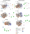
Docking results between NEST and different ligands. (A) Phosphinic acid. (B) PMSF. (C) CBDP. (D) Mipafox. (E) DFP. (F) PV. The ligands are shown in stick and surface of the protein is shown as surface and colored by hydrophobicity. Phosphinic acid and PMSF are non-aging inhibitors while CBDP, Mipafox, and DFP are aging compounds. PV is an artificial substrate of NTE. Dash lines with different colors in the 2D maps indicate different types of interactions, which are shown in the figure. Phe1066 is highlighted with a blue box.
Figure 7
Figure 7
Overlay of the docking results of NEST with non-aging and aging inhibitors. (A) Binding of three aging inhibitors CBDP (light red), Mipafox (pink), and DFP (dark red) with the NEST model. (B) Binding of two non-aging inhibitors PMSF (dark green), and phosphinic acid (light green) with the NEST model. The protein is shown in surface and colored by hydrophobicity. The phosphate groups of the inhibitors are colored in yellow and the alkane groups of the inhibitors are colored in gray.

Similar articles

References

    1. Kienesberger P.C., Oberer M., Lass A., Zechner R. Mammalian patatin domain containing proteins: A family with diverse lipolytic activities involved in multiple biological functions. J. Lipid Res. 2009;50:S63–S68. doi: 10.1194/jlr.R800082-JLR200. - DOI - PMC - PubMed
    1. UniProt UniProtKB—Q8IY17 (PLPL6_HUMAN) 2019. [(accessed on 26 November 2019)]. Available online: http://www.uniprot.org/uniprot/Q8IY17.
    1. Glynn P. Neuronal phospholipid deacylation is essential for axonal and synaptic integrity. Biochim. Biophys. Acta Mol. Cell Biol. Lipids. 2013;1831:633–641. doi: 10.1016/j.bbalip.2012.07.023. - DOI - PubMed
    1. Van Tienhoven M., Atkins J., Li Y., Glynn P. Human Neuropathy Target Esterase Catalyzes Hydrolysis of Membrane Lipids. J. Biol. Chem. 2002;277:20942–20948. doi: 10.1074/jbc.M200330200. - DOI - PubMed
    1. Zaccheo O., Dinsdale D., Meacock P.A., Glynn P. Neuropathy Target Esterase and Its Yeast Homologue Degrade Phosphatidylcholine to Glycerophosphocholine in Living Cells. J. Biol. Chem. 2004;279:24024–24033. doi: 10.1074/jbc.M400830200. - DOI - PubMed

Substances