

Azorean Black Tea (Camellia sinensis) Antidermatophytic and Fungicidal Properties
- PMID: 38067505
- PMCID: PMC10707949
- DOI: 10.3390/molecules28237775
Azorean Black Tea (Camellia sinensis) Antidermatophytic and Fungicidal Properties
Abstract
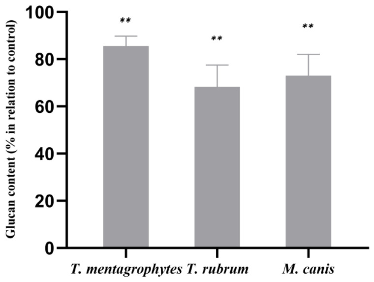
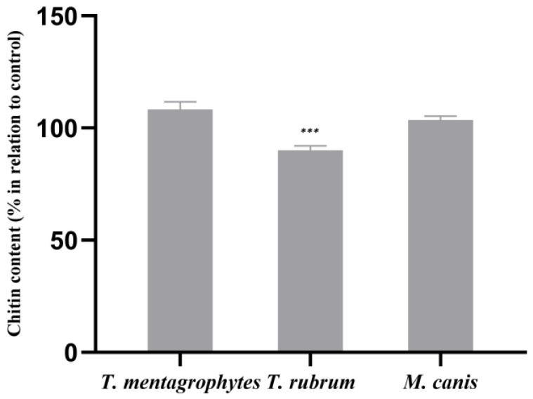
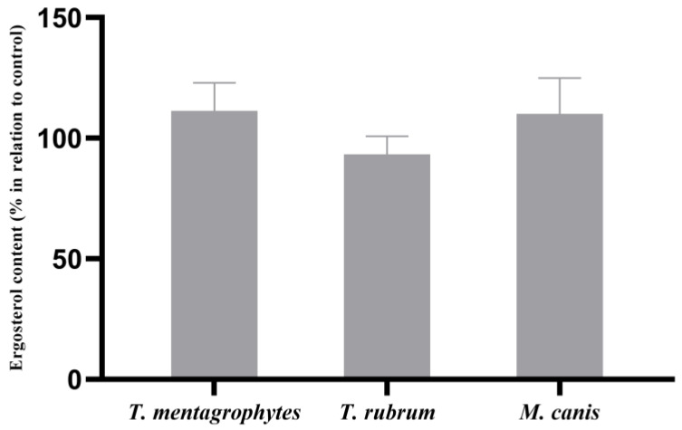
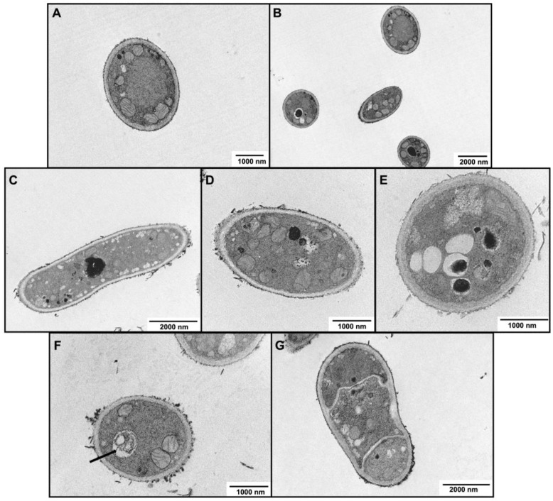
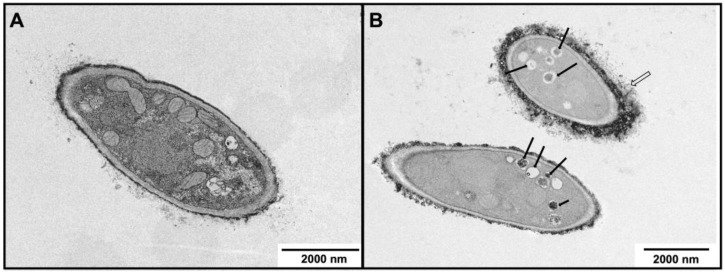
The treatment of dermatophytoses, the most common human fungal infections, requires new alternatives. The aim of this study was to determine the antidermatophytic activity of the aqueous Azorean Black Tea extract (ABT), together with an approach to the mechanisms of action. The phytochemical analysis of ABT extract was performed by HPLC. The dermatophytes susceptibility was assessed using a broth microdilution assay; potential synergies with terbinafine and griseofulvin were evaluated by the checkerboard assay. The mechanism of action was appraised by the quantification of the fungal cell wall chitin and β-1,3-glucan, and by membrane ergosterol. The presence of ultrastructural modifications was studied by Transmission Electron Microscopy (TEM). The ABT extract contained organic and phenolic acids, flavonoids, theaflavins and alkaloids. It showed an antidermatophytic effect, with MIC values of 250 µg/mL for Trichophyton mentagrophytes, 125 µg/mL for Trichophyton rubrum and 500 µg/mL for Microsporum canis; at these concentrations, the extract was fungicidal. An additive effect of ABT in association to terbinafine on these three dermatophytes was observed. The ABT extract caused a significant reduction in β-1,3-glucan content, indicating the synthesis of this cell wall component as a possible target. The present study identifies the antidermatophytic activity of the ABT and highlights its potential to improve the effectiveness of conventional topical treatment currently used for the management of skin or mucosal fungal infections.
Keywords: Azorean Black Tea; Camellia sinensis; antidermatophytic; antifungal; cell wall; chitin; dermatophytes; ergosterol; glucan.
Conflict of interest statement
The authors declare no conflict of interest.
Figures
Similar articles
-
In vitro and in vivo antidermatophytic activities of some Iranian medicinal plants.Med Mycol. 2015 Nov;53(8):852-9. doi: 10.1093/mmy/myv032. Epub 2015 Jun 19. Med Mycol. 2015. PMID: 26092105
-
Phytochemical composition of cedar tar of the atlas and it's in vitro antifungal activity against Trichophyton rubrum, Trichophyton mentagrophytes and Microsporum canis.Pak J Pharm Sci. 2024 Mar;37(2):257-263. Pak J Pharm Sci. 2024. PMID: 38767092
-
In vitro antidermatophytic activity and cytotoxicity of extracts derived from medicinal plants and marine algae.J Mycol Med. 2018 Sep;28(3):561-567. doi: 10.1016/j.mycmed.2018.07.001. Epub 2018 Jul 27. J Mycol Med. 2018. PMID: 30060991
-
How to: perform antifungal susceptibility testing of microconidia-forming dermatophytes following the new reference EUCAST method E.Def 11.0, exemplified by Trichophyton.Clin Microbiol Infect. 2021 Jan;27(1):55-60. doi: 10.1016/j.cmi.2020.08.042. Epub 2020 Sep 8. Clin Microbiol Infect. 2021. PMID: 32916260 Review.
-
The Emerging Terbinafine-Resistant Trichophyton Epidemic: What Is the Role of Antifungal Susceptibility Testing?Dermatology. 2022;238(1):60-79. doi: 10.1159/000515290. Epub 2021 May 31. Dermatology. 2022. PMID: 34058736 Review.
Cited by
-
VdAHA1 positively regulate pathogenicity in Verticillium dahliae.Front Microbiol. 2025 May 26;16:1535187. doi: 10.3389/fmicb.2025.1535187. eCollection 2025. Front Microbiol. 2025. PMID: 40491833 Free PMC article.
-
Caffeine Protects Keratinocytes from Trichophyton mentagrophytes Infection and Behaves as an Antidermatophytic Agent.Int J Mol Sci. 2024 Jul 30;25(15):8303. doi: 10.3390/ijms25158303. Int J Mol Sci. 2024. PMID: 39125871 Free PMC article.
-
Secondary Metabolites with Antioxidant and Antimicrobial Activities from Camellia fascicularis.Curr Issues Mol Biol. 2024 Jul 2;46(7):6769-6782. doi: 10.3390/cimb46070404. Curr Issues Mol Biol. 2024. PMID: 39057046 Free PMC article.
-
The role of VdSti1 in Verticillium dahliae: insights into pathogenicity and stress responses.Front Microbiol. 2024 Apr 4;15:1377713. doi: 10.3389/fmicb.2024.1377713. eCollection 2024. Front Microbiol. 2024. PMID: 38638896 Free PMC article.
References
MeSH terms
Substances
Grants and funding
- CENTRO-01-0145-FEDER- 000012- HealthyAging2020 and CENTRO-01-0145-FEDER- 022095: ViraVector; the COMPETE 2020 - Operational Programme for Competitiveness and Internation-alisation/European Regional Development Fund (ERDF)
- UIDB/04539/2020 and UIDP/04539/2020/Portuguese national funds via FCT - Fundação para a Ciência e a Tecnolo-gia,I.P.
LinkOut - more resources
Full Text Sources
Medical
