

Resistomycin Suppresses Prostate Cancer Cell Growth by Instigating Oxidative Stress, Mitochondrial Apoptosis, and Cell Cycle Arrest
- PMID: 38067602
- PMCID: PMC10708360
- DOI: 10.3390/molecules28237871
Resistomycin Suppresses Prostate Cancer Cell Growth by Instigating Oxidative Stress, Mitochondrial Apoptosis, and Cell Cycle Arrest
Abstract
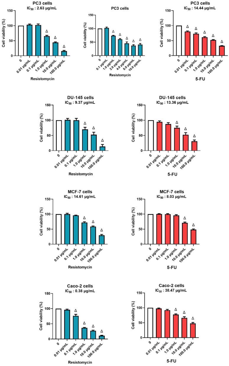
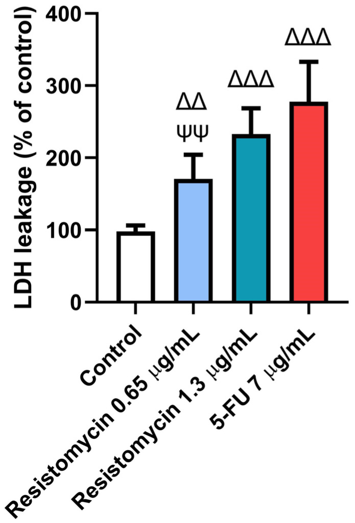
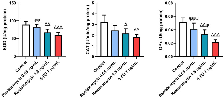
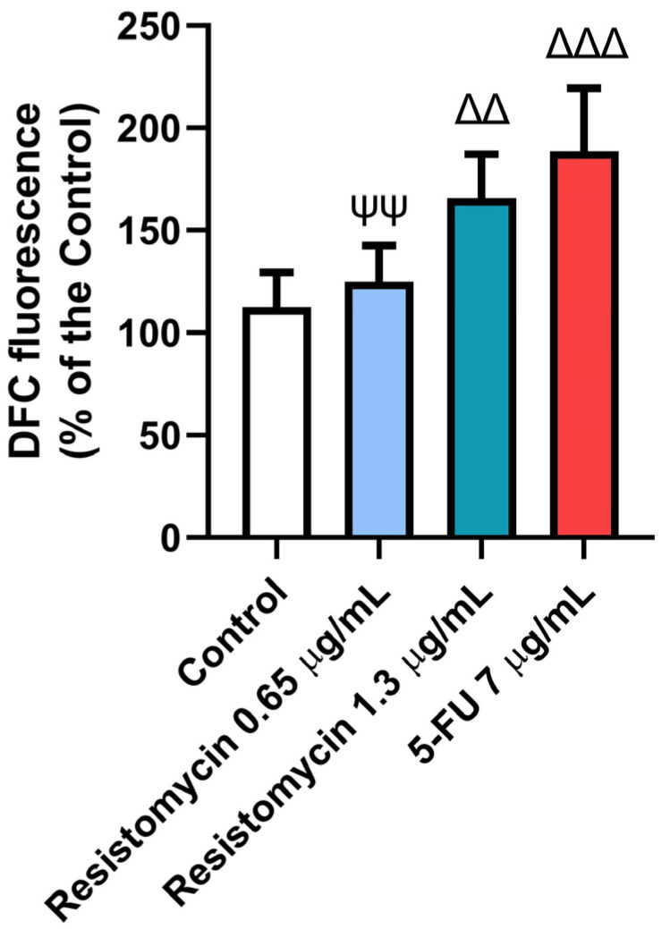
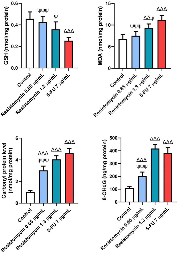
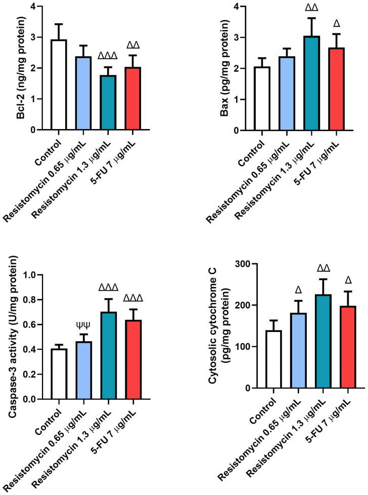
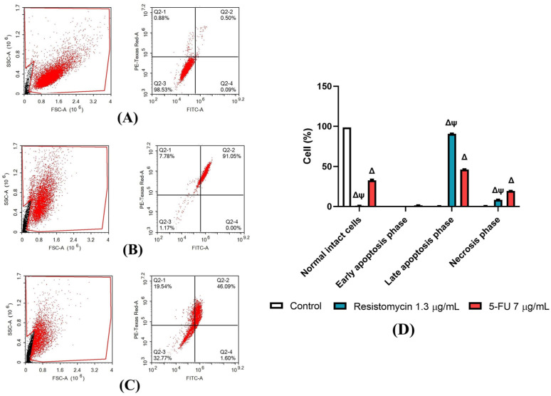
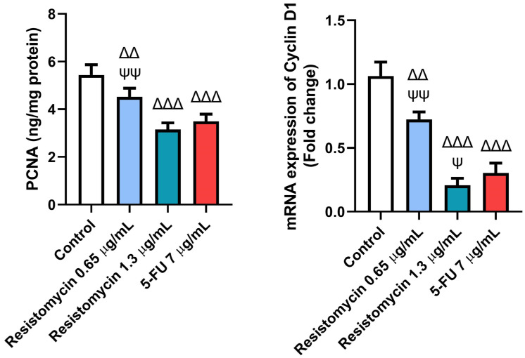
Globally, prostate cancer is among the most threatening and leading causes of death in men. This study, therefore, aimed to search for an ideal antitumor strategy with high efficacy, low drug resistance, and no or few adverse effects. Resistomycin is a natural antibiotic derived from marine actinomycetes, and it possesses various biological activities. Prostate cancer cells (PC3) were treated with resistomycin (IC12.5: 0.65 or IC25: 1.3 µg/mL) or 5-fluorouracil (5-FU; IC25: 7 µg/mL) for 24 h. MTT assay and flow cytometry were utilized to assess cell viability and apoptosis. Oxidative stress, apoptotic-related markers, and cell cycle were also assessed. The results revealed that the IC50 of resistomycin and 5-FU on PC3 cells were 2.63 µg/mL and 14.44 µg/mL, respectively. Furthermore, treated cells with the high dose of resistomycin showed an increased number of apoptotic cells compared to those treated with the lower dose. Remarkable induction of reactive oxygen species generation and lactate dehydrogenase (LDH) leakage with high malondialdehyde (MDA), carbonyl protein (CP), and 8-hydroxyguanosine (8-OHdG) contents were observed in resistomycin-treated cells. In addition, marked declines in glutathione (GSH), superoxide dismutase (SOD), catalase (CAT), and glutathione peroxidase (GPx) in PC3 cells subjected to resistomycin therapy were observed. Resistomycin triggered observable cell apoptosis by increasing Bax, caspase-3, and cytosolic cytochrome c levels and decreasing Bcl-2 levels. In addition, notable downregulation of proliferating cell nuclear antigen (PCNA) and cyclin D1 was observed in resistomycin-treated cancerous cells. According to this evaluation, the antitumor potential of resistomycin, in a concentration-dependent manner, in prostate cancer cells was achieved by triggering oxidative stress, mitochondrial apoptosis, and cell cycle arrest in cancer cells. In conclusion, our investigation suggests that resistomycin can be considered a starting point for developing new chemotherapeutic agents for human prostate cancer.
Keywords: 5-FU; apoptosis; oxidative stress; prostate cancer; resistomycin.
Conflict of interest statement
The authors declare no conflict of interest.
Figures
Similar articles
-
Suppression of human prostate cancer cell growth by ciprofloxacin is associated with cell cycle arrest and apoptosis.Int J Oncol. 2003 Apr;22(4):787-94. Int J Oncol. 2003. PMID: 12632069
-
Piper nigrum ethanolic extract rich in piperamides causes ROS overproduction, oxidative damage in DNA leading to cell cycle arrest and apoptosis in cancer cells.J Ethnopharmacol. 2016 Aug 2;189:139-47. doi: 10.1016/j.jep.2016.05.020. Epub 2016 May 10. J Ethnopharmacol. 2016. PMID: 27178634
-
Metformin and thymoquinone co-treatment enhance 5-fluorouracil cytotoxicity by suppressing the PI3K/mTOR/HIF1α pathway and increasing oxidative stress in colon cancer cells.Biofactors. 2023 Jul-Aug;49(4):831-848. doi: 10.1002/biof.1947. Epub 2023 Mar 16. Biofactors. 2023. PMID: 36929658
-
The variable chemotherapeutic response of Malabaricone-A in leukemic and solid tumor cell lines depends on the degree of redox imbalance.Phytomedicine. 2015 Jul 15;22(7-8):713-23. doi: 10.1016/j.phymed.2015.05.007. Epub 2015 May 29. Phytomedicine. 2015. PMID: 26141757
-
Effects of astaxanthin on oxidative stress induced by Cu2+ in prostate cells.J Zhejiang Univ Sci B. 2017 Feb.;18(2):161-171. doi: 10.1631/jzus.B1500296. J Zhejiang Univ Sci B. 2017. PMID: 28124844 Free PMC article.
Cited by
-
The Potential Anticancer Potency of Kolaviron on Colorectal Adenocarcinoma (Caco-2) Cells.Anticancer Agents Med Chem. 2024;24(15):1097-1108. doi: 10.2174/0118715206288807240527165444. Anticancer Agents Med Chem. 2024. PMID: 38835121
-
RBM15B Promotes Prostate Cancer Cell Proliferation via PCNA m6A Modification.Cell Biochem Biophys. 2025 Mar;83(1):1237-1248. doi: 10.1007/s12013-024-01558-5. Epub 2024 Oct 3. Cell Biochem Biophys. 2025. PMID: 39361104
-
[Multi-omics analysis of the relationship between oxidative stress-related gene and prostate cancer].Beijing Da Xue Xue Bao Yi Xue Ban. 2025 Aug 18;57(4):633-643. doi: 10.19723/j.issn.1671-167X.2025.04.002. Beijing Da Xue Xue Bao Yi Xue Ban. 2025. PMID: 40754897 Free PMC article. Chinese.
References
-
- Majid M., Farhan A., Baig M.W., Khan M.T., Kamal Y., Hassan S.S.U., Bungau S., Haq I.-U. Ameliorative Effect of Structurally Divergent Oleanane Triterpenoid, 3-Epifriedelinol from Ipomoea batatas against BPA-Induced Gonadotoxicity by Targeting PARP and NF-κB Signaling in Rats. Molecules. 2023;28:290. doi: 10.3390/molecules28010290. - DOI - PMC - PubMed
MeSH terms
Substances
Grants and funding
LinkOut - more resources
Full Text Sources
Medical
Research Materials
Miscellaneous
