

Helium Cold Atmospheric Plasma Causes Morphological and Biochemical Alterations in Candida albicans Cells
- PMID: 38067648
- PMCID: PMC10707892
- DOI: 10.3390/molecules28237919
Helium Cold Atmospheric Plasma Causes Morphological and Biochemical Alterations in Candida albicans Cells
Abstract
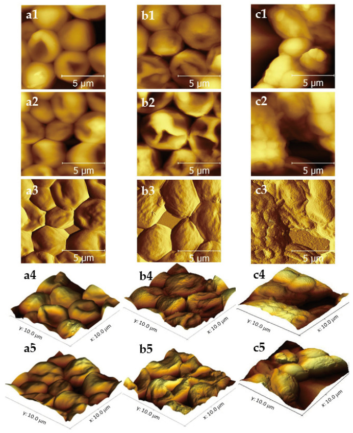
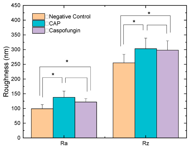
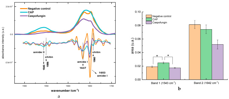
(1) Background: Previous studies reported the promising inhibitory effect of cold atmospheric plasma (CAP) on Candida albicans. However, the exact mechanisms of CAP's action on the fungal cell are still poorly understood. This study aims to elucidate the CAP effect on C. albicans cell wall, by evaluating the alterations on its structure and biochemical composition; (2) Methods: C. albicans cells treated with Helium-CAP were analyzed by atomic force microscopy (AFM) and Fourier transform infrared spectroscopy (FTIR) in order to detect morphological, topographic and biochemical changes in the fungal cell wall. Cells treated with caspofungin were also analyzed for comparative purposes; (3) Results: Expressive morphological and topographic changes, such as increased roughness and shape modification, were observed in the cells after CAP exposure. The alterations detected were similar to those observed after the treatment with caspofungin. The main biochemical changes occurred in polysaccharides content, and an overall decrease in glucans and an increase in chitin synthesis were detected; (4) Conclusions: Helium-CAP caused morphological and topographic alterations in C. albicans cells and affected the cell wall polysaccharide content.
Keywords: AFM; Candida albicans; Caspofungin; FTIR; chitin; cold atmospheric plasma; glucans.
Conflict of interest statement
The authors declare no conflict of interest.
Figures


Similar articles
-
Caspofungin-induced β(1,3)-glucan exposure in Candida albicans is driven by increased chitin levels.mBio. 2023 Aug 31;14(4):e0007423. doi: 10.1128/mbio.00074-23. Epub 2023 Jun 28. mBio. 2023. PMID: 37377417 Free PMC article.
-
Nanoscale effects of caspofungin against two yeast species, Saccharomyces cerevisiae and Candida albicans.Antimicrob Agents Chemother. 2013 Aug;57(8):3498-506. doi: 10.1128/AAC.00105-13. Epub 2013 May 13. Antimicrob Agents Chemother. 2013. PMID: 23669379 Free PMC article.
-
Paradoxical growth of Candida albicans in the presence of caspofungin is associated with multiple cell wall rearrangements and decreased virulence.Antimicrob Agents Chemother. 2014;58(2):1071-83. doi: 10.1128/AAC.00946-13. Epub 2013 Dec 2. Antimicrob Agents Chemother. 2014. PMID: 24295973 Free PMC article.
-
Caspofungin resistance in Candida albicans: genetic factors and synergistic compounds for combination therapies.Braz J Microbiol. 2022 Sep;53(3):1101-1113. doi: 10.1007/s42770-022-00739-9. Epub 2022 Mar 29. Braz J Microbiol. 2022. PMID: 35352319 Free PMC article. Review.
-
Natural products targeting the synthesis of β(1,3)-D-glucan and chitin of the fungal cell wall. Existing drugs and recent findings.Phytomedicine. 2021 Jul 15;88:153556. doi: 10.1016/j.phymed.2021.153556. Epub 2021 Mar 27. Phytomedicine. 2021. PMID: 33958276 Review.
Cited by
-
Effect of cold atmospheric plasma on common oral pathogenic microorganisms: a narrative review.Ann Med. 2025 Dec;57(1):2457518. doi: 10.1080/07853890.2025.2457518. Epub 2025 Jan 27. Ann Med. 2025. PMID: 39865862 Free PMC article. Review.
References
-
- Nogueira M.F., Istel F., Jenull S., Walker L.A., Gow N., Lion T. Quantitative Analysis of Candida Cell Wall Components by Flow Cytometry with Triple-Fluorescence Staining. J. Microbiol. Mod. Technol. 2017;2:1–9. doi: 10.15744/2575-5498.2.101. - DOI
-
- Perez-Nadales E., Almeida Nogueira M.F., Baldin C., Castanheira S., El Ghalid M., Grund E., Lengeler K., Marchegiani E., Mehrotra P.V., Moretti M., et al. Fungal Model Systems and the Elucidation of Pathogenicity Determinants. Fungal Genet. Biol. 2014;70:42–67. doi: 10.1016/j.fgb.2014.06.011. - DOI - PMC - PubMed
MeSH terms
Substances
LinkOut - more resources
Full Text Sources
Miscellaneous
