. 2023 Dec 4;28(23):7926.
doi: 10.3390/molecules28237926.

Pharmacological Evidence That Dictyostelium Differentiation-Inducing Factor 1 Promotes Glucose Uptake Partly via an Increase in Intracellular cAMP Content in Mouse 3T3-L1 Cells

Affiliations

Pharmacological Evidence That Dictyostelium Differentiation-Inducing Factor 1 Promotes Glucose Uptake Partly via an Increase in Intracellular cAMP Content in Mouse 3T3-L1 Cells

Yuzuru Kubohara et al. Molecules. .

Abstract

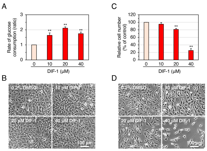
Differentiation-inducing factor 1 (DIF-1) isolated from the cellular slime mold Dictyostelium discoideum can inhibit mammalian calmodulin-dependent cAMP/cGMP phosphodiesterase (PDE1) in vitro. DIF-1 also promotes glucose uptake, at least in part, via a mitochondria- and AMPK-dependent pathway in mouse 3T3-L1 fibroblast cells, but the mechanism underlying this effect has not been fully elucidated. In this study, we investigated the effects of DIF-1 on intracellular cAMP and cGMP levels, as well as the effects that DIF-1 and several compounds that increase cAMP and cGMP levels have on glucose uptake in confluent 3T3-L1 cells. DIF-1 at 20 μM (a concentration that promotes glucose uptake) increased the level of intracellular cAMP by about 20% but did not affect the level of intracellular cGMP. Neither the PDE1 inhibitor 8-methoxymethyl-3-isobutyl-1-methylxanthine at 10-200 μM nor the broad-range PDE inhibitor 3-isobutyl-1-methylxanthine at 40-400 μM had any marked effects on glucose uptake. The membrane-permeable cAMP analog 8-bromo-cAMP at 200-1000 μM significantly promoted glucose uptake (by 20-25%), whereas the membrane-permeable cGMP analog 8-bromo-cGMP at 3-100 μM did not affect glucose uptake. The adenylate cyclase activator forskolin at 1-10 μM promoted glucose uptake by 20-30%. Thus, DIF-1 may promote glucose uptake by 3T3-L1 cells, at least in part, via an increase in intracellular cAMP level.

Keywords: DIF-1; Dictyostelium discoideum; PDE1; cAMP; diabetes; forskolin; obesity.


Conflict of interest statement

The authors declare no conflict of interest.

Figures

Figure 1
Figure 1
(A) Chemical structure of DIF-1. (B) Proposed scheme for the mechanisms of action of DIF-1. DIF-1–mediated uncoupling of mitochondrial activities may disrupt ATP production and activate AMP kinase, which may then induce GLUT1 translocation to the plasma membrane (PM) and glucose uptake [23,24]. Glucose may be metabolized immediately via glycolysis and via the TCA cycle [24,25]. DIF-1 may also inhibit PDE1 [26] and thereby increase intracellular cAMP and/or cGMP levels, which might promote glucose uptake [29,30].
Figure 2
Figure 2
Effects of DIF-1 on glucose uptake (A,B) and growth of 3T3-L1 cells (C,D). (A) Confluent 3T3-L1 cells were incubated for 16–20 h in the presence of 0.2% dimethyl sulfoxide (DMSO; vehicle) or 10–40 μM DIF-1, and the rate of glucose consumption was assessed. The data are mean ± SD of three independent experiments. ** p < 0.01 versus control (by t-test). Note that the rate of glucose consumption measured by our method matches well with that of glucose uptake assessed with 2-[1,2-3H]deoxy-d-glucose [23]; therefore, sometimes we refer to ‘glucose consumption’ as ‘glucose uptake’. (B) Confluent 3T3-L1 cells were incubated as in (A) and observed under a phase-contrast microscope; representative photos are shown. (C) Growing 3T3-L1 cells were incubated for 3 days in the presence of 0.2% DMSO (vehicle) or 10–40 μM DIF-1, and relative cell number was determined. The data are mean ± SD of three independent experiments. ** p < 0.01 versus control (by t-test). (D) Growing 3T3-L1 cells were incubated for 3 days as in (C) and observed under a phase-contrast microscope; representative photos are shown.
Figure 3
Figure 3
Effects of DIF-1 on intracellular cAMP and cGMP contents in confluent 3T3-L1 cells. Cells were incubated in the presence of 20 μM DIF-1, harvested at the indicated time points, and assayed for (A) cAMP or (B) cGMP content. The data are mean ± SD of triplicate samples in a single experiment. ** p < 0.01 versus Time 0 control (by t-test).
Figure 4
Figure 4
(A) Effects of 8-MIBMX and DIF-1 on glucose uptake in confluent 3T3-L1 cells. Cells were incubated for 16–20 h in the presence of 0.2% or 0.4% DMSO (vehicle) and the indicated concentrations of 8-MIBMX or DIF-1, and the rate of glucose consumption was assessed. The data are mean ± SD of three independent experiments. * p < 0.05, ** p < 0.01 versus control (by t-test). (B) Combinatorial effects of 8-MIBMX and DIF-1 on glucose uptake. Cells were incubated for 16–20 h in the presence of 0.3% DMSO (vehicle) and the indicated concentrations of 8-MIBMX, DIF-1, or both, and the rate of glucose consumption was assessed. The data are mean ± SD of four independent experiments. ** p < 0.01 versus control (by t-test); n.s., not significant (by ANOVA).
Figure 5
Figure 5
(A) Effects of IBMX and DIF-1 on glucose uptake in confluent 3T3-L1 cells. Cells were incubated for 16–20 h in the presence of 0.2% DMSO (vehicle) and the indicated concentrations of IBMX or DIF-1, and the rate of glucose consumption was assessed. The data are mean ± SD of three independent experiments. * p < 0.01 versus control (by t-test). (B) Combinatorial effects of IBMX and DIF-1 on glucose uptake. Cells were incubated for 16–20 h in the presence of 0.2% DMSO (vehicle) and the indicated concentrations of MIBMX, DIF-1, or both, and the rate of glucose consumption was assessed. The data are mean ± SD of four independent experiments. * p < 0.01 versus control (by t-test); n.s., not significant (by ANOVA).
Figure 6
Figure 6
(A) Effects of Br-cAMP and DIF-1 on glucose uptake in confluent 3T3-L1 cells. Cells were incubated for 16–20 h in the presence of 0.1% DMSO (vehicle) and the indicated concentrations of Br-cAMP or DIF-1, and the rate of glucose consumption was assessed. The data are mean ± SD of three independent experiments. * p < 0.05, ** p < 0.01 versus control (by t-test). (B) Combinatorial effects of Br-cAMP and DIF-1 on glucose uptake. Cells were incubated for 16–20 h in the presence of 0.1% DMSO (vehicle) and the indicated concentrations of Br-cAMP, DIF-1, or both, and the rate of glucose consumption was assessed. The data are mean ± SD of five independent experiments. ** p < 0.01 versus control (by t-test); # p < 0.05 (by ANOVA).
Figure 7
Figure 7
(A) Effects of Br-cGMP and DIF-1 on glucose uptake in confluent 3T3-L1 cells. Cells were incubated for 16–20 h in the presence of 0.1% DMSO (vehicle) and the indicated concentrations of Br-cGMP or DIF-1, and the rate of glucose consumption was assessed. The data are mean ± SD of three independent experiments. ** p < 0.01 versus control (by t-test). (B) Combinatorial effects of Br-cGMP and DIF-1 on glucose uptake. Cells were incubated for 16–20 h in the presence of 0.1% DMSO (vehicle) and the indicated concentrations of Br-cGMP, DIF-1, or both, and the rate of glucose consumption was assessed. The data are mean ± SD of three independent experiments. ** p < 0.01 versus control (by t-test); n.s., not significant (by ANOVA).
Figure 8
Figure 8
(A) Effects of forskolin and DIF-1 on glucose uptake in confluent 3T3-L1 cells. Cells were incubated for 16–20 h in the presence of 0.2% DMSO (vehicle) and the indicated concentrations of forskolin or DIF-1, and the rate of glucose consumption was assessed. The data are mean ± SD of three independent experiments. * p < 0.05, ** p < 0.01 versus control (by t-test). (B) Combinatorial effects of forskolin and DIF-1 on glucose uptake. Cells were incubated for 16–20 h in the presence of 0.2% DMSO (vehicle) and the indicated concentrations of forskolin, DIF-1, or both, and the rate of glucose consumption was assessed. The data are mean ± SD of four independent experiments. * p < 0.05, ** p < 0.01 versus control (by t-test); # p < 0.05 (by ANOVA).
Figure 9
Figure 9
(A) Effects of DNP and DIF-1 on glucose uptake in confluent 3T3-L1 cells. Cells were incubated for 16–20 h in the presence of 0.1% DMSO (vehicle) and the indicated concentrations of DNP or DIF-1, and the rate of glucose consumption was assessed. The data are mean ± SD of three independent experiments. ** p < 0.01 versus control (by t-test). (B) Combinatorial effects of DNP and Br-cAMP on glucose uptake. Cells were incubated for 16–20 h in the presence of 0.1% DMSO (vehicle) and the indicated concentrations of DNP and/or Br-cAMP, and the rate of glucose consumption was assessed. The data are mean ± SD of four independent experiments. * p < 0.05, ** p < 0.01 versus control (by t-test); ## p < 0.01 (by ANOVA).
Figure 10
Figure 10
Effects of the compounds affecting cAMP/cGMP levels on the growth of 3T3-L1 (A) and HeLa cells (B). Growing cells were incubated for 3 days in the presence of the indicated compounds, and relative cell number was determined. All media contained 0.2% (black bars) or 0.4% (gray bars) DMSO (vehicle). The data are mean ± SD of three independent experiments. * p < 0.05; ** p < 0.01 versus control (by t-test).
Figure 11
Figure 11
Proposed scheme for the action of DIF-1 in 3T3-L1 cells. DIF-1 promotes glucose uptake, in part via a mitochondria- and AMPK-dependent pathway [23,24] and possibly also via an increase in intracellular cAMP level (this study). The mitochondrial uncoupler DNP can stimulate the mitochondria- and AMPK-dependent pathway and thereby promote GLUT1 translocation to the plasma membrane (PM) and glucose uptake [24], whereas Br-cAMP (membrane-permeable analog of cAMP) and forskolin (adenylate cyclase activator) increase the intracellular cAMP level and thus mimic the effect of DIF-1 (this study). Note that an increase in intracellular cAMP has been shown to induce GLUT1 translocation to the plasma membrane and promote glucose uptake in 3T3-L1 adipocytes and brown fat cells [29,30].

Similar articles

Cited by

References

    1. Konijn T.M., van de Meene J.G.C., Bonner J.T., Barkley D.S. The acrasin activity of adenosine-3’,5’-cyclic phosphate. Proc. Natl. Acad. Sci. USA. 1967;58:1152–1154. doi: 10.1073/pnas.58.3.1152. - DOI - PMC - PubMed
    1. Darmon M., Brachet P., Pereira da Silva L.H. Chemotactic signals induce cell differentiation in Dictyostelium discoideum. Proc. Natl. Acad. Sci. USA. 1975;72:3163–3166. doi: 10.1073/pnas.72.8.3163. - DOI - PMC - PubMed
    1. Devreotes P. Dictyostelium discoideum: A model system for cell-cell interactions in development. Science. 1989;245:1054–1058. doi: 10.1126/science.2672337. - DOI - PubMed
    1. Morris H.R., Taylor G.W., Masento M.S., Jermyn K.A., Kay R.R. Chemical structure of the morphogen differentiation inducing factor from Dictyostelium discoideum. Nature. 1987;328:811–814. doi: 10.1038/328811a0. - DOI - PubMed
    1. Kay R.R., Berks M., Traynor D. Morphogen hunting in Dictyostelium discoideum. Development. 1989;107:81–90. doi: 10.1242/dev.107.Supplement.81. - DOI - PubMed

Substances

LinkOut - more resources