

Red Blood Cells' Area Deformation as the Origin of the Photoplethysmography Signal
- PMID: 38067889
- PMCID: PMC10708758
- DOI: 10.3390/s23239515
Red Blood Cells' Area Deformation as the Origin of the Photoplethysmography Signal
Abstract
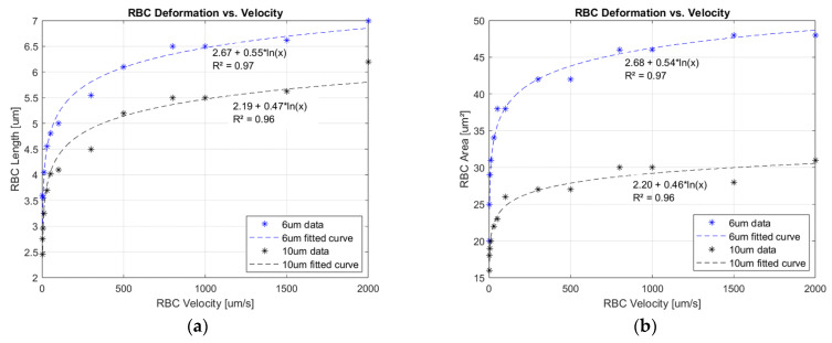
The origin of the photoplethysmography (PPG) signal is a debatable topic, despite plausible models being addressed. One concern revolves around the correlation between the mechanical waveform's pulsatile nature and the associated biomechanism. The interface between these domains requires a clear mathematical or physical model that can explain physiological behavior. Describing the correct origin of the recorded optical waveform not only benefits the development of the next generation of biosensors but also defines novel health markers. In this study, the assumption of a pulsatile nature is based on the mechanism of blood microcirculation. At this level, two interconnected phenomena occur: variation in blood flow velocity through the capillary network and red blood cell (RBC) shape deformation. The latter effect was qualitatively investigated in synthetic capillaries to assess the experimental data needed for PPG model development. Erythrocytes passed through 10 µm and 6 µm microchannel widths with imposed velocities between 50 µm/s and 2000 µm/s, according to real scenarios. As a result, the length and area deformation of RBCs followed a logarithmic law function of the achieved traveling speeds. Applying radiometric expertise on top, mechanical-optical insights are obtained regarding PPG's pulsatile nature. The mathematical equations derived from experimental data correlate microcirculation physiologic with waveform behavior at a high confidence level. The transfer function between the biomechanics and the optical signal is primarily influenced by the vasomotor state, capillary network orientation, concentration, and deformation performance of erythrocytes.
Keywords: mathematical transfer function; microcirculation; photoplethysmography origin; red blood cell shape deformation; vasomotor activity.
Conflict of interest statement
The authors declare no conflict of interest.
Figures


References
-
- Bonsmann M.R. Blutdruckversuche an der Maus und Ratte mittels Photozelle. Naunyn-Schmiedebergs Arch. Exp. Pathol. Und Pharmakol. 1934;176:460–467. doi: 10.1007/BF01930644. - DOI
-
- Hertzman A.B. Photoelectric Plethysmography of the Fingers and Toes in Man. Exp. Biol. Med. 1937;37:529–534. doi: 10.3181/00379727-37-9630. - DOI
-
- Hertzman A.B., Dillon J.B. Distinction between arterial, venous and flow components in photoelectric plethysmography. Am. Physiol. Soc. 1940;130:177–185. doi: 10.1152/ajplegacy.1940.130.1.177. - DOI
MeSH terms
Grants and funding
LinkOut - more resources
Full Text Sources
Research Materials
