. 2023 Nov 24;12(23):7297.
doi: 10.3390/jcm12237297.

Local MRI before and after Tumor Resection in Neuroblastoma: Impact of Residual Disease on Event Free Survival

Affiliations

Local MRI before and after Tumor Resection in Neuroblastoma: Impact of Residual Disease on Event Free Survival

Jürgen F Schäfer et al. J Clin Med. .

Abstract

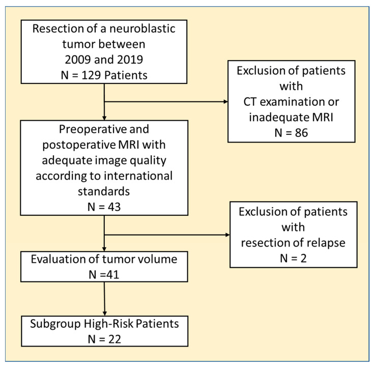
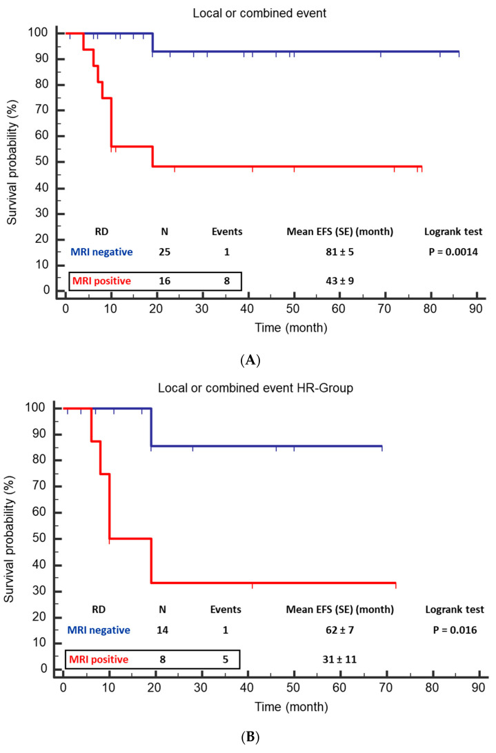
(1) Background: The study aimed to investigate the influence of MRI-defined residual disease on local tumor control after resection of neuroblastic tumors in patients without routine adjuvant radiotherapy. (2) Methods: Patients, who underwent tumor resection between 2009 and 2019 and received a pre- and postoperative MRI, were included in this retrospective single-center study. Measurement of residual disease (RD) was performed using standardized criteria. Primary endpoint was the local or combined (local and metastatic) event free survival (EFS). (3) Results: Forty-one patients (20 female) with median age of 39 months were analyzed. Risk group analysis showed eleven low-, eight intermediate-, and twenty-two high-risk patients (LR, IR, HR). RD was found in 16 cases by MRI. A local or combined relapse or progression was found in nine patients of whom eight patients had RD (p = 0.0004). From the six patients with local or combined relapse in the HR group, five had RD (p = 0.005). Only one of 25 patients without RD had a local event. Mean EFS (month) was significantly higher if MRI showed no residual tumor (81 ± 5 vs. 43 ± 9; p = 0.0014) for the total cohort and the HR subgroup (62 ± 7 vs. 31 ± 11; p = 0.016). (4) Conclusions: In our series, evidence of residual tumor, detectable by MRI, was associated with insufficient local control, resulting in relapses or local progression in 50% of patients. Only one of the patients without residual tumor had a local relapse.

Keywords: EFS; MRI; high-risk neuroblastoma; irradiation; residual tumor; surgical resection.


Conflict of interest statement

The authors declare no conflicts of interest.

Figures

Figure 1
Figure 1
The graph shows the workflow of patient selection and the evaluation of the entire cohort as well as the subgroup of high-risk patients.
Figure 2
Figure 2
Example of imaging analysis in a 15-year-old male patient with ganglioneuroblastoma. (AE) Before and (FJ) after resection. (A,F) T2 weighted images with fat saturation. (B,G) Non-enhanced T1 weighted images and (D,I) after application of contrast medium. (C,H) DWI with high b-value (ADC map not shown). (E,J) The multiplanar reconstruction respective 3D volume analysis; the tumor volume is colored green, and in the volume rendering method it is colored light yellow. Before resection, the tumor located anterior to the spine reached the left renal hilum and had extensions into the ipsilateral neuroforamina. MRI characterized the tumor as inhomogeneous with hemorrhage (star), inhomogeneous to even absent contrast enhancement. There was a moderate diffusion restriction in the area of the non-regressively altered tumor parts. Tumor volume was 234 mL. After resection, two extensions could be detected unchanged in the neuroforamina and a small RD adjacent to the aorta. The total volume was 11 mL. According to the surgical report, the extensions into the neuroforamina were not resected (IME) because this patient did not have a high-risk situation. There was no tumor progression.
Figure 3
Figure 3
Example of a case with residual tumor and local progression in a 10-year-old girl with ganglioneuroblastoma, N-MYC non-amplified tumor, INSS Stage IV. The T2 weighted images are shown. (A,B) Before resection, (C,D) after resection, and E and F during follow-up. (A,C,E) Transversal images at the level of the primary tumor on the left suprarenal side. After surgery and in the follow-up, no tumor can be detected (C,E). Enlarged left lymph node before surgery (B) and after surgery (D) (arrows). According to the surgical report, a CME was performed. In the course of 12 months, the tumor progressed (F) (arrow).
Figure 4
Figure 4
Event free survival (EFS) of patients with positive MRI for residual disease vs. negative MRI. (A) For the total cohort, mean EFS was 46 months (CI 95% 30 to 61) for positive MRI and 79 months (CI 95% 70 to 88; p = 0.0035) for negative MRI. (B) In the HR group, mean EFS was 36 months (CI 95% 16 to 56) vs. 63 months (CI 95% 51 to 75).

References

    1. Ries L.A.G., Smith M.A., Gurney J., Linet M., Tamra T., Young J., Bunin G. Cancer Incidence and Survival among Children and Adolescents: United States SEER Program 1975–1995. National Cancer Institute; Rockville, MD, USA: 1999.
    1. Maris J.M. Recent advances in neuroblastoma. N. Engl. J. Med. 2010;362:2202–2211. doi: 10.1056/NEJMra0804577. - DOI - PMC - PubMed
    1. Maris J.M., Hogarty M.D., Bagatell R., Cohn S.L. Neuroblastoma. Lancet. 2007;369:2106–2120. doi: 10.1016/S0140-6736(07)60983-0. - DOI - PubMed
    1. Bar-Sever Z., Biassoni L., Shulkin B., Kong G., Hofman M.S., Lopci E., Manea I., Koziorowski J., Castellani R., Boubaker A., et al. Guidelines on nuclear medicine imaging in neuroblastoma. Eur. J. Nucl. Med. Mol. Imaging. 2018;45:2009–2024. doi: 10.1007/s00259-018-4070-8. - DOI - PubMed
    1. Brisse H.J., McCarville M.B., Granata C., Krug K.B., Wootton-Gorges S.L., Kanegawa K., Giammarile F., Schmidt M., Shulkin B.L., Matthay K.K., et al. Guidelines for imaging and staging of neuroblastic tumors: Consensus report from the International Neuroblastoma Risk Group Project. Radiology. 2011;261:243–257. doi: 10.1148/radiol.11101352. - DOI - PubMed

LinkOut - more resources