Review
. 2023 Nov 27;12(23):7349.
doi: 10.3390/jcm12237349.

Pathomechanisms of Diabetic Kidney Disease

Affiliations
Review

Pathomechanisms of Diabetic Kidney Disease

Satyesh K Sinha et al. J Clin Med. .

Abstract

The worldwide occurrence of diabetic kidney disease (DKD) is swiftly rising, primarily attributed to the growing population of individuals affected by type 2 diabetes. This surge has been transformed into a substantial global concern, placing additional strain on healthcare systems already grappling with significant demands. The pathogenesis of DKD is intricate, originating with hyperglycemia, which triggers various mechanisms and pathways: metabolic, hemodynamic, inflammatory, and fibrotic which ultimately lead to renal damage. Within each pathway, several mediators contribute to the development of renal structural and functional changes. Some of these mediators, such as inflammatory cytokines, reactive oxygen species, and transforming growth factor β are shared among the different pathways, leading to significant overlap and interaction between them. While current treatment options for DKD have shown advancement over previous strategies, their effectiveness remains somewhat constrained as patients still experience residual risk of disease progression. Therefore, a comprehensive grasp of the molecular mechanisms underlying the onset and progression of DKD is imperative for the continued creation of novel and groundbreaking therapies for this condition. In this review, we discuss the current achievements in fundamental research, with a particular emphasis on individual factors and recent developments in DKD treatment.

Keywords: hemodynamic; inflammatory and fibrotic factors; metabolic; mineralocorticoid receptor; osteopontin; renin angiotensin aldosterone system; targeted therapies.


Conflict of interest statement

S.B.N. reports grant funding from: NIH/NCATS, NIH/NIMHD, CDC, Travere, Bayer; Editorial Board: American Journal of Kidney Disease; Associate Editor, Journal of the American Society of Nephrology; Advisory Board/Steering Committee: Boehringer Ingelheim/Lilly Pharmaceuticals, AstraZeneca, Bayer, NovoNordisk, Janssen Pharmaceutical; Consultant/National Leader: Bayer-ND-CKD; Honoraria: 2021–2022, 2023 Hypertension Highlights, ADA, 2023 ASN nephSAP. S.K.S. has no conflict of interest.

Figures

Figure 1
Figure 1
Factors that promote diabetes and DKD. The diagram illustrates that the progression of DKD encompasses a range of factors, including metabolic, hemodynamic, inflammatory, fibrotic processes, and genetic and epigenetic factors. These components impact various pathways that oversee intricate intracellular signaling networks, ultimately resulting in both functional and structural alterations within the kidney. Abbreviations: DKD: diabetic kidney disease, MR: mineralocorticoid receptor, AGE: advanced glycation end products, ACE: angiotensin-converting enzyme, RAAS: renin–angiotensin–aldosterone system, TGF-β: transforming growth factor-β, MAPKs: mitogen-activated protein kinases, PI3K/AKT: phosphatidylinositol-3-kinase/Ak strain transforming, JAK/STAT: Janus kinase/signal transducers and activators of transcription.
Figure 2
Figure 2
Role of metabolic factors in the development of DKD. The diagram depicts the impact of metabolic disruptions in hyperglycemia on various pathways, namely PKC, AGE, hexosamine, and the polyol pathway. The PKC pathway is linked to heightened production of ECM, especially collagen IV, leading to the expansion of mesangial cells and the development of glomerulosclerosis. The interaction between AGE and RAGE triggers downstream signaling molecules, including MAPK, p38, SAPK/JNK, ERK1/2, and JAK/STAT. The hexosamine pathway is responsible for generating glucosamine-6-p, which in turn promotes the release of cytokines such as TGF-β, ICAM-1, VCAM-1, TNF-α, CTGF, and PAI-1. The polyol pathway results in a redox imbalance between NADH and NAD+ in diabetes mellitus. Collectively, these processes contribute to the progression of DKD. Abbreviations: DKD: diabetic kidney disease, glucosamine-6-P: glucosamine-6-phosphate, PKC: protein kinase C, AGE: advanced glycation end products, ECM: extracellular matrix, RAGE: receptor for advanced glycation end products, SAPK/JNK: stress-activated protein kinases/Jun amino-terminal kinases, ERK1/2: extracellular signal-regulated kinase 1/2, JAK/STAT: Janus kinase/signal transducers and activators of transcription, TGF-β: transforming growth factor- β, ICAM-1: intercellular adhesion molecule 1, VCAM-1: vascular cell adhesion molecule 1, TNF-α: tumor necrosis factor alpha, CTGF: connective tissue growth factor, PAI-1: plasminogen activator inhibitor 1.
Figure 3
Figure 3
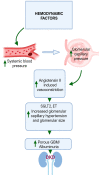
Role of hemodynamic factors in the development of DKD. The figure demonstrates the process by which diabetes gives rise to hemodynamic changes, characterized by elevated systemic blood pressure and heightened intraglomerular pressure. These factors collectively encourage angiotensin II-induced vasoconstriction. The amplified glomerular capillary pressure and increased glomerular size led to a more permeable GBM, consequently resulting in albuminuria. These combined effects contribute to the development of DKD. Abbreviations: SGLT2: sodium–glucose cotransporter 2, ET: endothelin, DKD: diabetic kidney disease, GBM: glomerular basement membrane.
Figure 4
Figure 4
Role of inflammatory factors in the development of DKD. The diagram illustrates how inflammation plays a role in the development of DKD. In diabetes, there is an infiltration and activation of immune cells, including macrophages, T-cells, and B-cells, within renal tissue. This leads to an increased expression of proinflammatory cytokines like IL-1, IL-6, IL-18, and TNF-α. Consequently, this intensifies the inflammatory responses within the renal tissue, contributing to its damage. Abbreviations: DKD: diabetic kidney disease, IL: interleukin, TNF-α: tumor necrosis factor alpha, MR: mineralocorticoid receptor, ROS: reactive oxygen species, Mφ: macrophage, DC: dendritic cell.
Figure 5
Figure 5
Role of fibrotic factors in the development of DKD. The diagram depicts the involvement of renal fibrosis in the development of DKD. This fibrotic process encompasses various pathways, including TGF-β, MAPK, Wnt/β-catenin, PI3K/AKT, JAK/STAT, and NOTCH signaling. Within the TGF-β signaling pathway, Smad 2/3 mediates the production of ECM. The MAPK pathway leads to an increase in signaling transduction mediated by P38MAPK, ERK1/2, and JNK, resulting in elevated collagen IV levels. The Wnt/β-catenin pathway contributes to podocyte loss and mesangial cell apoptosis through the regulation of WT-1-associated genes and the cleavage of cas-3 and PARP, respectively. The collective action of the PI3K/AKT-JAK/STAT-NOTCH signaling pathways promotes PI3K/AKT, STAT1/3, and Snail signaling, leading to the induced expression of fibronectin/collagen IV, TGF-β1, VEGF, ACE, α-SMA, MMP2/9. Ultimately, these molecules drive the process of renal fibrosis in DKD. Abbreviations: DKD: diabetic kidney disease, WT-1: Wilms tumor-1, a master regulator of gene expression in podocytes, TGF-β: transforming growth factor-beta, MAPK: mitogen-activated protein kinases, ERK1/2: extracellular signal-regulated kinase 1/2, JNK: c-Jun amino-terminal kinase, Cas-3: caspase-3, PARP: polyADP-ribose polymerase, PI3K/AKT: phosphatidylinositol-3-kinase/Ak strain transforming, STAT1/3: signal transducer and activator of transcription 1/3, JAK/STAT: Janus kinase/signal transducers and activators of transcription, Smad 2/3: suppressor of mothers against decapentaplegic 2/3, ECM: extracellular matrix, VEGF: vascular endothelial growth factor, ACE: angiotensin-converting enzyme, a-SMA: alpha smooth muscle actin, MMP2/9: matrix metalloproteinase2/9.
Figure 6
Figure 6
Mineralocorticoid receptor (MR) and aldosterone in DKD. The diagram depicts the involvement of the MR and aldosterone in the development of DKD. The MR functions as an intracellular receptor, instigating the inflammatory cascade with aldosterone by generating ROS in the mitochondria, a process further amplified by Rac1. Both Rac1 and aldosterone contribute to fibrosis and inflammation by activating inflammasomes, NLRP3. The activation of the NLRP3 inflammasome, results in renal inflammation, fibrosis, and glomerular sclerosis. Abbreviations: DKD: diabetic kidney disease, MR: mineralocorticoid receptor, ROS: reactive oxygen species NLRP3: nucleotide-binding domain, leucine-rich-containing family, pyrin domain-containing-3.
Figure 7
Figure 7
OPN-mediated fibrosis in DKD. The diagram illustrates OPN-mediated signaling pathways. The detrimental impacts of OPN occur via its interaction with receptors, integrins, and CD44. Binding to these receptors leads to significant proinflammatory functions, enabling OPN to trigger the activation of various pathways, including cell survival, cell proliferation, angiogenesis, migration, and fibrosis. Abbreviations: OPN: osteopontin, ntOPN: N-terminal osteopontin, TGF-β: transforming growth factor-β, ERK/MAPK: extracellular signal-regulated kinase/mitogen-activated protein kinase, JNK/MAPK: jun N-terminal kinase/mitogen-activated protein kinase, PI3K/AKT/mTOR: phosphatidylinositol-3-kinase/Ak strain transforming/mammalian target of rapamycin, FAK/ERK1/2/NF-κB: focal adhesion kinase/extracellular signal-regulated kinase 1 and 2/nuclear factor kappa B.
Figure 8
Figure 8
Epigenetics in DKD. Histone modifications, specifically acetylation and methylation, along with DNA methylation, are linked to the aberrant regulation of genes associated with inflammation and fibrosis in DKD. Abbreviations: DM: diabetes mellitus, DKD: diabetic kidney disease, Me: methylation, Ac: acetylation, DNA: deoxyribonucleic acid.

References

    1. Guariguata L., Whiting D.R., Hambleton I., Beagley J., Linnenkamp U., Shaw J.E. Global estimates of diabetes prevalence for 2013 and projections for 2035. Diabetes Res. Clin. Pract. 2014;103:137–149. doi: 10.1016/j.diabres.2013.11.002. - DOI - PubMed
    1. Cho N.H., Shaw J.E., Karuranga S., Huang Y., da Rocha Fernandes J.D., Ohlrogge A.W., Malanda B. IDF Diabetes Atlas: Global estimates of diabetes prevalence for 2017 and projections for 2045. Diabetes Res. Clin. Pract. 2018;138:271–281. doi: 10.1016/j.diabres.2018.02.023. - DOI - PubMed
    1. Tuttle K.R., Bakris G.L., Bilous R.W., Chiang J.L., de Boer I.H., Goldstein-Fuchs J., Hirsch I.B., Kalantar-Zadeh K., Narva A.S., Navaneethan S.D., et al. Diabetic kidney disease: A report from an ADA Consensus Conference. Diabetes Care. 2014;37:2864–2883. doi: 10.2337/dc14-1296. - DOI - PMC - PubMed
    1. Chen Y., Lee K., Ni Z., He J.C. Diabetic Kidney Disease: Challenges, Advances, and Opportunities. Kidney Dis. 2020;6:215–225. doi: 10.1159/000506634. - DOI - PMC - PubMed
    1. Gheith O., Farouk N., Nampoory N., Halim M.A., Al-Otaibi T. Diabetic kidney disease: World wide difference of prevalence and risk factors. J. Nephropharmacol. 2016;5:49–56. doi: 10.4103/1110-9165.197379. - DOI - PMC - PubMed