Review
. 2023 Nov 21;12(23):3921.
doi: 10.3390/plants12233921.

Arabinogalactan-Proteins as Boron-Acting Enzymes, Cross-Linking the Rhamnogalacturonan-II Domains of Pectin

Affiliations
Review

Arabinogalactan-Proteins as Boron-Acting Enzymes, Cross-Linking the Rhamnogalacturonan-II Domains of Pectin

Rifat Ara Begum et al. Plants (Basel). .

Abstract

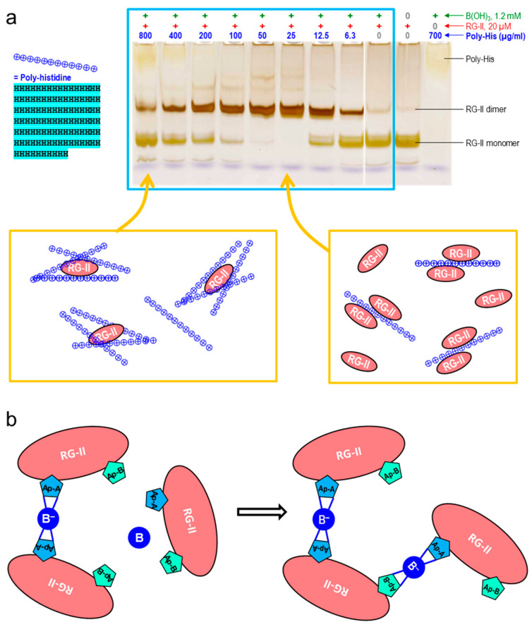
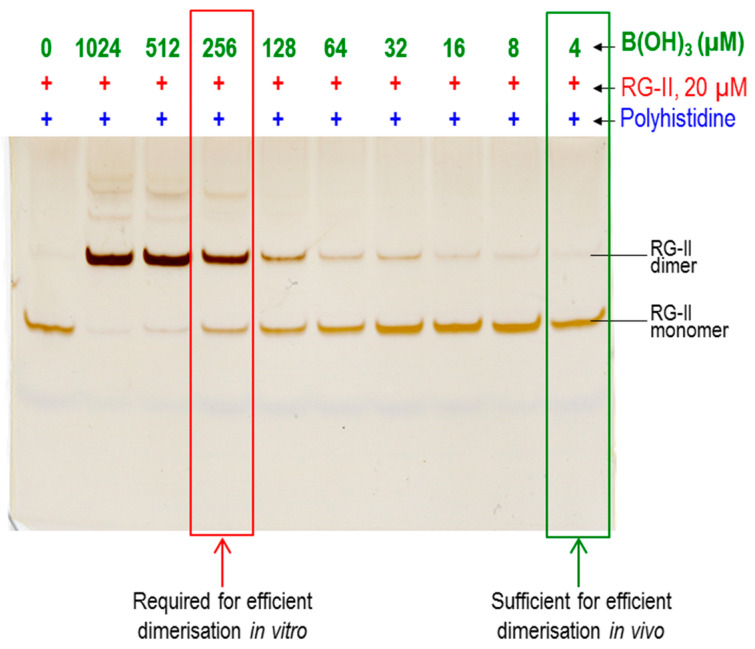
Most pectic rhamnogalacturonan-II (RG-II) domains in plant cell walls are borate-bridged dimers. However, the sub-cellular locations, pH dependence, reversibility and biocatalyst involvement in borate bridging remain uncertain. Experiments discussed here explored these questions, utilising suspension-cultured plant cells. In-vivo pulse radiolabelling showed that most RG-II domains dimerise extremely quickly (<4 min after biosynthesis, thus while still intraprotoplasmic). This tallies with the finding that boron withdrawal causes cell wall weakening within 10-20 min, and supports a previously proposed biological role for boron/RG-II complexes specifically at the wall/membrane interface. We also discuss RG-II monomer ↔ dimer interconversion as monitored in vitro using gel electrophoresis and a novel thin-layer chromatography method to resolve monomers and dimers. Physiologically relevant acidity did not monomerise dimers, thus boron bridge breaking cannot be a wall-loosening mechanism in 'acid growth'; nevertheless, recently discovered RG-II trimers and tetramers are unstable and may thus underpin reversible wall loosening. Dimerising monomers in vitro by B(OH)3 required the simultaneous presence of RG-II-binding 'chaperones': co-ordinately binding metals and/or ionically binding cationic peptides. Natural chaperones of the latter type include highly basic arabinogalactan protein fragments, e.g., KHKRKHKHKRHHH, which catalyse a reaction [2 RG-II + B(OH)3 → RG-II-B-RG-II], suggesting that plants can 'enzymically' metabolise boron.

Keywords: Ca2+; Pb2+; acid-growth; arabinogalactan-proteins; borate diesterase; boron; chaperones; pectin; rhamnogalacturonan-II; trimers of RG-II.


Conflict of interest statement

The authors declare no conflict of interest.

Figures

Figure 1
Figure 1
Primary structure of rhamnogalacturonan-II (RG-II) (adapted from references [18,19,20]). Abbreviations: Ac (in a circle), O-acetyl ester group; AceA, l-aceric acid; Api, d-apiose; Araf, l-arabinofuranose; Arap, l-arabinopyranose; DHA, 3-deoxy-d-lyxo-heptulosaric acid; Gal, galactose (d- or l-, as stated on the diagram); GalA, d-galacturonic acid; GlcA, d-glucuronic acid; KDO, 3-deoxy-d-manno-octulosonic acid; Me (in a circle), methyl ester group (attached to position 6, so that the uronic acid residue is not anionic); MeFuc, 2-O-methyl-l-fucose; MeGalA, d-galacturonic acid with methyl ether group(s) possibly on the 3- and/or 4-positions [20]; MeXyl, 2-O-methyl-d-xylose; Rha, l-rhamnose. All sugar residues are in the pyranose ring form unless indicated ‘f’ for furanose. The numerals (2, 3, 3′, 4, 5) indicate the position to which the neighbouring sugar residue makes a glycosidic bond; this bond is from the anomeric carbon (i.e., from C-1 of most sugars but from C-2 of the ketoses (DHA and KDO)). Arrows represent glycosidic bonds. Residues shaded red carry a negative charge at physiological pH (two negative charges in the case of DHA); those shaded green or purple are non-ionic; those shaded purple are the sites of boron binding. The grey rectangle represents the homogalacturonan-like backbone of RG-II.
Figure 2
Figure 2
Boron bridging of RG-II. (a) Schematic representation of the B-bridging of two RG-II molecules [24]. The two sugar chains shown in black are portions of the backbones (effectively, short homogalacturonans) of these two RG-IIs. A side chain ‘A’ is attached via its apiose residue to each backbone, and a tetrahedral B atom (shown in red) can bond to two apiose residues, forming borate diesters and thereby dimerising the RG-II. (b) Effect of pH on peptide-mediated RG-II dimerisation, as revealed by polyacrylamide gel electrophoresis [23]. Eight reaction mixtures each contained 16 µM RG-II monomer, 1.2 mM boric acid, a 50 mM buffer, and a low concentration of a chaperone (0.55 µM polyhistidine). The pH values of the reaction mixtures are shown above the gel. After incubation at 20 °C for 16 h, the products were analysed by PAGE and stained with silver. The main dimer bands are highlighted by the white box. The putative RG-II trimer (Tr) and tetramer (Te) are also labelled. The marker mixture (MM, lane 1) contained 0.8 µg each of RG-II monomer (M) and dimer (D). The histogram shows the calculated effect of pH on the net charge of the polyhistidine (positive charges, blue bars) and RG-II (negative charges, red bars). Adapted with permission from Ref. [23], ©2023, Wiley Blackwell. (c) Thin-layer chromatography (TLC) as an alternative to PAGE for resolving monomers from dimers (unpublished). The samples loaded were (left to right) (i) oligogalacturonides, (ii) RG-II monomer, (iii) a mixture of RG-II dimer plus RG-I. The plastic-backed TLC plate was Merck silica gel. The plate was pre-washed sequentially by gentle rocking in acetone/acetic acid/water (1:1:1, 30 min, to remove thymol-stainable material from the plate), acetone (×2, each 15 min, to remove the acetic acid), 2% pyridine in acetone (15 min, to neutralise residual traces of acidity), and acetone (15 min, to remove traces of pyridine and expedite drying), dried, then baked in an oven for 30 min at 120 °C. The aqueous samples were loaded, then thoroughly dried in a stream of air (room temperature), and the chromatogram was developed (same day) in freshly prepared butan-1-ol/acetic acid/water (13:10:17 by vol.). The separated bands were stained in thymol/H2SO4 [21]. Previously unpublished work. Abbreviation: DP, degree of polymerisation.
Figure 3
Figure 3
The essential reaction involved in boron bridging of RG-II [22]. Part of the neutral apiose residue of side chain A (in each of two monomeric RG-II molecules) is shown. The charges of its four near-neighbouring anionic sugar residues are rendered as red circles (two α-GalA residues in the RG-II backbone plus an α-GalA and a β-GalA residue attached to the rhamnose adjacent to the apiose). Monomeric RG-II has a total of about 11 additional anionic sugar residues, giving it a net charge of about −14 at the pH (4.8) used in our experiments. It is evident that the close approach of two RG-II monomers for B bridging requires the overcoming of considerable electrostatic repulsion; in addition, dimerisation introduces an additional negative charge on the previously neutral B atom (Adapted with permission from Ref. [22], ©2022, Portland Press).
Figure 4
Figure 4
14C-Labelling of monomeric and dimeric RG-II domains in suspension-cultured rose and arabidopsis cells [52]. A pulse of [14C]glucose, supplied at time 0, was consumed by the cells within ~1–2 h, during which time the radioactivity was incorporated into cell wall polysaccharides including RG-II. At intervals, the monomeric and dimeric RG-II domains were assayed for 14C. Incorporation of 14C into the monomer (triangles) occurred during the first 2 h, and then almost stopped. Dimer radiolabelling (circles) mirrored this closely during the first 2 h, indicating that any given newly synthesised RG-II domain was highly likely to become dimerised almost immediately. But between 2 and 17 h, during which interval the vast majority of the radiolabelled cohort of molecules would have been in the cell wall, some additional dimer labelling continued, indicating slight RG-II dimerisation within the cell wall. Adapted with permission from Ref. [52], ©2022, Oxford University Press).
Figure 5
Figure 5
Inorganic cations as RG-II chaperones [23]. Each reaction mixture initially contained 16 µM pure monomeric RG-II and a 50 mM buffer suitable for the pH indicated above the gel lanes. (a) With no additional components; (b) with added 1.2 mM boric acid but no divalent cations; (c) with 100 µM Pb2+ but no B(OH)3; (d) with 5000 µM Ca2+ but no B(OH)3; (e) with 4 µM Pb2+ plus 1.2 mM boric acid; (f) with 2000 µM Ca2+ plus 1.2 mM boric acid. After 16 h incubation, the products were analysed by PAGE, resolving monomeric and dimeric RG-II. The blue ellipse in (f) highlights the (weak) ability of calcium to produce the dimer. Note that Pb2+ is >1000× more effective than Ca2+. MM, marker mixture of monomeric and dimeric RG-II for reference. Adapted with permission from Ref. [23], ©2023, Wiley Blackwell.
Figure 6
Figure 6
Polyhistidine as a cationic chaperone, catalysing the boron bridging of RG-II. (a) Each reaction mixture contained 100 µg/mL RG-II monomer (≈20 µM), 1.2 mM boric acid, 0–800 µg/mL (≈0–44 µM) polyhistidine (chloride salt), and 50 mM acetate (Na+) buffer pH 4.8. Controls (outside the blue rectangle) lacked boric acid or RG-II, as indicated above the gels. After 16 h, 0.8 µg of the RG-II was analysed by PAGE followed by silver staining [22]. Below the gel (orange rectangles) is an interpretation of why an optimum polyhistidine concentration exists: at 25 µg/mL, there is an excess of RG-II molecules, which are thus obliged to ‘share’ a polyhistidine molecule bringing them close enough together for dimerisation, whereas at 800 µg/mL, there is an excess of polyhistidine and thus little chance that two RG-II molecules will be brought close together on a single polyhistidine molecule [Gel image adapted with permission from Ref. [22], ©2022, Portland Press]. (b) Forming a trimer from a dimer plus a monomer requires at least 1 B bridge to be made through the apiose residue of RG-II’s side chain “B” (Ap-B) rather than through the preferred Ap-A. The trimers and tetramers are unstable: this makes them difficult to isolate, but marks them out as being of biological interest as potentially reversible B bridges. B in a blue circle represents boric acid; B in a blue circle represents a tetravalent boron atom.
Figure 7
Figure 7
(Above) The amino acid sequence of AGP31, with the N-terminal signal peptide removed. The sequence is aligned to emphasise the regular spacing of positively charged lysine (K) and occasional arginine (R) residues. In the native molecule, many of the proline residues (P) are hydroxyproline, and many of those are glycosylated [56,57]. Also shown is the remarkably histidine-rich domain (cyan). Amino acids with charged side-chains are colour-coded as follows: cations in shades of blue (His, cyan; Lys, blue; Arg, dark blue) and anions in shades of red (Asp, pink; Glu, red). The sequence is taken from NCBI database (https://www.ncbi.nlm.nih.gov/protein/OAP15690.1; accessed on 5 September 2022 [58]) [adapted with permission from Ref. [22], ©2022, Portland Press]. (Below) Four remarkably basic oligopeptides which appear in arabidopsis arabinogalactan proteins AGP17, 18, 19 and 31.
Figure 8
Figure 8
Effect of boron concentration on the polyhistidine-catalysed dimerisation of RG-II [22]. Reaction mixtures initially contained 50 µg/mL monomeric RG-II (20 µM) with 0–1024 µM boric acid and 50 mM acetate (Na+, pH 4.8), with polyhistidine (chloride salt; 50 µg/mL; 2.8 µM). After 4 h at 20 °C, the RG-II was analysed by PAGE. Note the difference in B(OH)3 concentration required for effective dimerisation in vitro (>250 µM) and that sufficing in the medium of cultured Rosa cells (3.3 µM). Adapted with permission from Ref. [22], ©2022, Portland Press.
Figure 9
Figure 9
The predominant glycosyl inositol phosphoryl ceramide (GIPC) species found in membranes of Rosa cell suspension cultures. Purple dot, potential site of binding of borate to three –OH groups. Green, glycosyl inositol moiety; red, long-chain 2-hydroxy-fatty acid; blue, long-chain base; dashed grey box, phytoceramide moiety. The structure was elucidated by mass spectrometric and thin-layer chromatographic analysis of Rosa GIPCs [11]. Abbreviations: Man, d-mannose, GlcA, d-glucuronic acid. Adapted with permission from Ref. [11], ©2014, Wiley Blackwell.
Figure 10
Figure 10
RG-II domains fixed in position by ‘egg-boxed’ homogalacturonan. (a) Suitably for B bridging of the RG-IIs. (b) unsuitably for B bridging.

Similar articles

Cited by

References

    1. Warington K. The effect of boric acid and borax on the broad bean and certain other plants. Ann. Bot. 1923;37:629–672. doi: 10.1093/oxfordjournals.aob.a089871. - DOI
    1. Rowe R.I., Eckhert C.D. Boron is required for zebrafish embryogenesis. J. Exp. Biol. 1999;202:1649–1654. doi: 10.1242/jeb.202.12.1649. - DOI - PubMed
    1. Glime J.M. Nutrient relations: Requirements and sources. In: Glime J.M., editor. Bryophyte Ecology. Volume 1. Michigan Technological University and the International Association of Bryologists; Houghton, MI, USA: 2017. pp. 1–32.
    1. Matsunaga T., Ishii T., Matsumoto S., Higuchi M., Darvill A., Albersheim P., O’Neill M.A. Occurrence of the primary cell wall polysaccharide rhamnogalacturonan II in pteridophytes, lycophytes, and bryophytes. Implications for the evolution of vascular plants. Plant Physiol. 2004;134:339–351. doi: 10.1104/pp.103.030072. - DOI - PMC - PubMed
    1. Chormova D., Messenger D.J., Fry S.C. Boron bridging of rhamnogalacturonan-II, monitored by gel electrophoresis, occurs during polysaccharide synthesis and secretion but not post-secretion. Plant J. 2014;77:534–546. doi: 10.1111/tpj.12403. - DOI - PMC - PubMed

LinkOut - more resources