. 2023 Nov 25;24(23):16758.
doi: 10.3390/ijms242316758.

miR-877-5p as a Potential Link between Triple-Negative Breast Cancer Development and Metabolic Syndrome

Affiliations

miR-877-5p as a Potential Link between Triple-Negative Breast Cancer Development and Metabolic Syndrome

Juana Moro et al. Int J Mol Sci. .

Abstract

Metabolic syndrome (MS) is a risk factor for breast cancer (BC) that increases its aggressiveness and metastasis. The prevalence of MS is higher in triple-negative breast cancer (TNBC), which is the molecular subtype with the worst prognosis. The molecular mechanisms underlying this association have not been fully elucidated. MiRNAs are small, non-coding RNAs that regulate gene expression. Aberrant expression of miRNAs in both tissues and fluids are linked to several pathologies. The aim of this work was to identify circulating miRNAs in patients with alterations associated with MS (AAMS) that also impact on BC. Using microarray technology, we detected 23 miRNAs altered in the plasma of women with AAMS that modulate processes linked to cancer. We found that let-7b-5p and miR-28-3p were decreased in plasma from patients with AAMS and also in BC tumors, while miR-877-5p was increased. Interestingly, miR-877-5p expression was associated with lower patient survival, and its expression was higher in PAM50 basal-like BC tumors compared to the other molecular subtypes. Analyses from public databases revealed that miR-877-5p was also increased in plasma from BC patients compared to plasma from healthy donors. We identified IGF2 and TIMP3 as validated target genes of miR-877-5p whose expression was decreased in BC tissue and moreover, was negatively correlated with the levels of this miRNA in the tumors. Finally, a miRNA inhibitor against miR-877-5p diminished viability and tumor growth of the TNBC model 4T1. These results reveal that miR-877-5p inhibition could be a therapeutic option for the treatment of TNBC. Further studies are needed to investigate the role of this miRNA in TNBC progression.

Keywords: breast cancer; metabolic syndrome; miRNAs.


Conflict of interest statement

The authors declare no conflict of interest. The funders had no role in the design of the study; in the collection, analyses, or interpretation of data; in the writing of the manuscript; or in the decision to publish the results.

Figures

Figure 1
Figure 1
The expression of circulating miRNAs with functions related to cancer was altered in the plasma of women with AAMS. (A) MiRNAs were isolated from plasma of women with AAMS or the control group and hybridized to GeneChip®miRNA 4.0 Array (Affymetrix) (n = 2, each sample was generated by pooling the plasma from nine women). Heatmap depicting the differentially expressed miRNAs (FSG < 0.05%, LogFC > 1. 5 and p value < 0.01) is shown. (B) Histogram depicting the KEGG pathways regulated by the validated target genes from these miRNAs determined through DIANA miRPath. The top 20 most significant pathways are shown. (C) Heatmap + showing the miRNAs and their KEGG pathways. Red arrows indicate KEGG pathways relevant to cancer and the miRNAs with stronger association to these pathways.
Figure 2
Figure 2
MiR-877-5p, -28-3p and let-7b-5p expression levels were altered in primary breast tumors from patients and their expression is dependent on the PAM50 subtype. (A) Expression levels of AAMS-modulated miRNAs were determined in primary tumors (PT) and adjacent normal tissue (ANT) of patients from TCGA BRCA data—Illumina HiSeq (n = 75). Significance was determined with paired t-test or Wilcoxon signed-rank test, when normality failed (*, p < 0.05). (B) Expression levels of miR-877-5p, miR-28-3p and let-7b-5p were determined in BC tissue from the different PAM50 molecular subtypes using TCGA BRCA data—Illumina HiSeq available in UCSC Xena tool (n = 1101). Basal means PAM50 basal-like BC; HER, HER2+ BC; Lum A, luminal A BC; Lum B, luminal B BC and Normal, normal-like BC. Significance was analyzed using one-way ANOVA or Kruskal–Wallis rank sum test, when normality failed. Asterisks indicate a significative difference compared to basal-like tumors (**, p < 0.01; ***, p < 0.001).
Figure 3
Figure 3
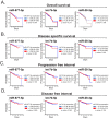
miR-877-5p expression was correlated with a diminished overall survival of patients. The impact of let-7b-5p, miR-28-3p and miR-877-5p in: (A) overall survival, (B) disease-specific survival, (C) progression-free interval and (D) disease-free interval of patients with BC was assessed in TCGA BRCA data—Illumina HiSeq using UCSC Xena tool. Log-rank test was carried out to determine significance.
Figure 4
Figure 4
MiR877-5p expression levels were increased in primary tumors from patients with PAM50 basal-like BC. Expression levels of miR-877-5p were determined in primary tumors (PT) and adjacent normal tissue (ANT) of patients with PAM50 basal-like BC from TCGA BRCA data—Illumina HiSeq. Significance was determined using Mann–Whitney test (balanced N = 41; **** p < 0.0001).
Figure 5
Figure 5
miR-877-5p expression was increased in plasma from patients with AAMS and from patients with BC. (A) miR-877-5 expression in plasma from women with AAMS or healthy donors (HD) (N = 18) was determined using stem loop RT-qPCR. Data were normalized to spike in cel-miR-39 and control group. Significance was evaluated using Mann–Whitney Wilcoxon signed-rank test (*, p < 0.05). (B) miR-877-5p expression in women with BC and HD was analyzed in the GSE73002 dataset (balanced N = 1280). Significance was evaluated using Mann–Whitney–Wilcoxon signed-rank test (***, p < 0.001).
Figure 6
Figure 6
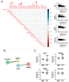
miR-877-5p expression levels were correlated with IGF2, ERBB2, TIMP3 and COL6A2 levels in mammary tumors. (A) Spearman test was performed to analyze correlation between miR-877-5p and its validated target genes in primary tumors of BC cohort TCGA BRCA data—Illumina HiSeq (n = 1101). Matrix correlation and dot plots of genes with low and moderate correlation are shown. Red arrows in the matrix indicate genes with low to moderate correlation with miR-877-5p. (B) Interaction network from STRING bioinformatic tool of the 4 genes with low to moderate correlation. Edges in black indicate co-expression; in green, text mining; and in blue, protein homology. The minimum required interaction score was 0.400 (medium confidence). (C) Expression levels of these genes in primary tumors (PT) and adjacent normal tissue (ANT) of patients from TCGA BRCA data—Illumina HiSeq (n = 75). Significance was determined using paired t-test or Wilcoxon signed-rank test, when normality failed (***, p < 0.001).
Figure 7
Figure 7
miR-877-5p inhibition decreased viability and adhesion in a model of TNBC. 4T1 cells were transfected with miR-877-5p inhibitor (877-5p inhibitor) or NC5 negative control (NC) (n = 3). (A) miR-877-5p expression was determined with RT-qPCR 24 h post-transfection or (B) 48 hours post-transfection. Data were normalized to U6 expression and control cells. (C) Twenty four hours post-transfection, cells were incubated with medium containing 1% FBS and cell viability was determined with MTS assay 48 h later. (D) Balb/c mice (n = 10) were inoculated with 4T1 cells s.c. On day 13, when the tumors were around 80 mm3, the animals were randomly divided and inoculated with PEI nanoparticles containing the miR-877-5p inhibitor or NC. Tumor size was determined with a digital caliper. The arrow indicates the day on which animals were treated with nanoparticles. Significance was determined using t-test (*, p < 0.05; ***, p < 0.001).
Figure 8
Figure 8
A cluster of circulating miRNAs dysregulated in plasma of women with AAMS has relevance in BC. Review diagram of the new insights presented in this paper. This diagram was performed using Canva free online resource (https://www.canva.com/ Accessed on 14 April 2023).

References

    1. Sung H., Ferlay J., Siegel R.L., Laversanne M., Soerjomataram I., Jemal A., Bray F. Global Cancer Statistics 2020: GLOBOCAN Estimates of Incidence and Mortality Worldwide for 36 Cancers in 185 Countries. CA Cancer J. Clin. 2021;71:209–249. doi: 10.3322/caac.21660. - DOI - PubMed
    1. Won K.A., Spruck C. Triple-negative Breast Cancer Therapy: Current and Future Perspectives. Int. J. Oncol. 2020;57:1245–1261. doi: 10.3892/ijo.2020.5135. - DOI - PMC - PubMed
    1. Yin L., Duan J.J., Bian X.W., Yu S.C. Triple-Negative Breast Cancer Molecular Subtyping and Treatment Progress. Breast Cancer Res. 2020;22:61. doi: 10.1186/s13058-020-01296-5. - DOI - PMC - PubMed
    1. Czene K., Lichtenstein P., Hemminki K. Environmental and Heritable Causes of Cancer among 9.6 Million Individuals in the Swedish Family-Cancer Database. Int. J. Cancer. 2002;99:260–266. doi: 10.1002/ijc.10332. - DOI - PubMed
    1. Willett W.C. Balancing Life-Style and Genomics Research for Disease Prevention. Science. 2002;296:695–698. doi: 10.1126/science.1071055. - DOI - PubMed

MeSH terms