. 2023 Dec 4;24(23):17104.
doi: 10.3390/ijms242317104.

Socrates: A Novel N-Ethyl-N-nitrosourea-Induced Mouse Mutant with Audiogenic Epilepsy

Affiliations

Socrates: A Novel N-Ethyl-N-nitrosourea-Induced Mouse Mutant with Audiogenic Epilepsy

Elena G Varlamova et al. Int J Mol Sci. .

Abstract

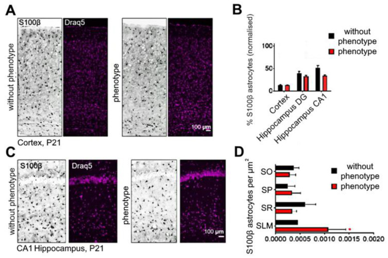
Epilepsy is one of the common neurological diseases that affects not only adults but also infants and children. Because epilepsy has been studied for a long time, there are several pharmacologically effective anticonvulsants, which, however, are not suitable as therapy for all patients. The genesis of epilepsy has been extensively investigated in terms of its occurrence after injury and as a concomitant disease with various brain diseases, such as tumors, ischemic events, etc. However, in the last decades, there are multiple reports that both genetic and epigenetic factors play an important role in epileptogenesis. Therefore, there is a need for further identification of genes and loci that can be associated with higher susceptibility to epileptic seizures. Use of mouse knockout models of epileptogenesis is very informative, but it has its limitations. One of them is due to the fact that complete deletion of a gene is not, in many cases, similar to human epilepsy-associated syndromes. Another approach to generating mouse models of epilepsy is N-Ethyl-N-nitrosourea (ENU)-directed mutagenesis. Recently, using this approach, we generated a novel mouse strain, soc (socrates, formerly s8-3), with epileptiform activity. Using molecular biology methods, calcium neuroimaging, and immunocytochemistry, we were able to characterize the strain. Neurons isolated from soc mutant brains retain the ability to differentiate in vitro and form a network. However, soc mutant neurons are characterized by increased spontaneous excitation activity. They also demonstrate a high degree of Ca2+ activity compared to WT neurons. Additionally, they show increased expression of NMDA receptors, decreased expression of the Ca2+-conducting GluA2 subunit of AMPA receptors, suppressed expression of phosphoinositol 3-kinase, and BK channels of the cytoplasmic membrane involved in protection against epileptogenesis. During embryonic and postnatal development, the expression of several genes encoding ion channels is downregulated in vivo, as well. Our data indicate that soc mutation causes a disruption of the excitation-inhibition balance in the brain, and it can serve as a mouse model of epilepsy.

Keywords: calcium ions; epileptiform activity; gene expression; mutagenesis; neurons; receptors; signaling.


Conflict of interest statement

The authors declare no conflict of interest.

Figures

Figure 1
Figure 1
The ratio of animals with epileptiform activity among offspring of the soc line of different backcross generations G3 (A) and G5–G6 (B).
Figure 2
Figure 2
Behavioral phenotyping of soc mice for the acoustic startle reaction (A), for general motor activity in the Mouse Open Field test (B), and when assessing orienting–exploratory activity (C) and learning ability in the CPAR test (D). Data are presented as mean ± SD. * significance of differences between the “phenotype” group and the “without phenotype” group (p < 0.05, Kolmogorov–Smirnov normality test and Mann–Whitney test).
Figure 3
Figure 3
Volcano plot of genes differentially expressed in socrates mouse brains (A). Log2 Fold change is plotted on the x-axis and the −log10 adjusted p value on the y-axis. Points are colored according to passing adjusted p value or adjusted p value and log2Fold Change filters. (B) The pseudo-Manhattan plot of significantly changed genes in soc mouse (phenotype) by chromosome.
Figure 4
Figure 4
ISH with RNA probes for the genes alpk1 (A), unc5d (B), zfp990 (C), slc17a6 (D), and pcp4l1 (E) on coronal cortical sections at P21.
Figure 5
Figure 5
Development of the neuronal network and differentiation of neurons isolated from “without phenotype” and “phenotype” soc mice. (A,B) Staining of cerebral cortex cells at 3 DIV (A) and 12 DIV (B) with the Calcein probe. (C,D) Changes in the expression of genes encoding proteins that regulate neuronal differentiation at 3 DIV (C) and 12 DIV (D). n/s—data not significant (p > 0.05), ** p < 0.01, and *** p < 0.001.
Figure 6
Figure 6
Characteristics of Ca2+ activity of neurons isolated from “without phenotype” and “phenotype” soc mice. (A,B) spontaneous Ca2+ signals of “without phenotype” neurons (A) and “phenotype” neurons (B). (C,D) Ca2+ signals of “without phenotype” (C) and “phenotype” neurons (D) when modeling epileptiform activity by excluding Mg2+ ions from the medium (Mg2+-free). (E,F) Ca2+ signals of “without phenotype” (E) and “phenotype” soc neurons (F) when modeling epileptiform activity using inhibition of GABA(A) receptors upon application of 10 μM of bicuculline. Typical Ca2+ signals of neurons are presented.
Figure 7
Figure 7
Analysis of the period (A) and amplitude (B) of Ca2+ impulses in “without phenotype” and “phenotype” soc neurons during spontaneous Ca2+ activity and modeling of epileptiform activity using the exclusion of Mg2+ ions (Mg2+-free) and inhibition of GABA(A)—receptors (bicuculline). Averaged results obtained on 4 cell cultures are presented. n/s—data not significant (p > 0.05), *** p < 0.001.
Figure 8
Figure 8
Expression of genes encoding isoforms of phosphoinositol 3-kinase and protein kinase C (A), inhibitory GABA receptors (GABA-A and GABA-B) (B), and excitatory glutamate receptors (AMPAR, NMDAR, and KAR) (C) in cultured cortical neurons obtained from “phenotype” soc mice: 1 (dashed line) is the level of gene expression in neurons obtained from “without phenotype” soc mice. n/s—data not significant (p > 0.05), * p < 0.05 and *** p < 0.001.
Figure 9
Figure 9
Secondary antibody fluorescence analysis reflecting phosphoinositol 3-kinase (A) protein levels, AMPAR (B), and NMDAR (C) subunits in “without phenotype” and “phenotype” soc neurons. The results presented in the figures correspond to Supplementary Figures S1 and S2. For each column, 300 ± 150 neurons were analyzed. * p < 0.05, ** p < 0.01, and *** p < 0.001.
Figure 10
Figure 10
Effect of AMPAR and NMDAR blockers on spontaneous Ca2+ activity in neurons isolated from soc mice. (A) Spontaneous Ca2+ signals of soc neurons. (B) Application of the AMPAR antagonist NBQX (10 mkM) against the background of spontaneous Ca2+ oscillations of soc neurons. (C) Application of the NMDAR antagonist D-AP5 (50 mkM) against the background of spontaneous Ca2+ oscillations of soc neurons. Typical Ca2+ signals of neurons in one experiment are presented.
Figure 11
Figure 11
Expression patterns of genes encoding protein kinases (A), GABA and glutamate receptors (B), and membrane ion pathways (C) in the nucleus of the brain of newborns (black bars) of soc mice and during their development at 1 month (red bars) and 1 year (blue bars). For 1—dotted line, the level of expression in the brain nucleus of “without phenotype” soc mice is achieved. n/s—data not significant (p > 0.05), * p < 0.05, ** p < 0.01, and *** p < 0.001.
Figure 12
Figure 12
Quantification of GABA+ interneurons in the cortex (A,B) and dentate gyrus (C,D). Representative images of P21 coronal sections. Data are presented as mean ± SD. ** significance of differences in the “phenotype” group compared to the “without phenotype” group (0.001 < ** p < 0.01, D’Agostino–Pearson normality test and Mann–Whitney test).
Figure 13
Figure 13
Quantification of the proportion of astrocytes in the cortex (A), the CA1 region of the hippocampus (C), considering different regions of the hippocampus (B) and dividing the CA1 region into zones (D). Representative images of P21 coronal sections. Data are presented as mean ± SD. * significance of differences between the “phenotype” and the “without phenotype” soc mice (0.01< * p < 0.05, D’Agostino–Pearson normality test and Mann–Whitney test).
Figure 14
Figure 14
Crossing scheme with backcross stage employed.

References

    1. Hill R.S., Walsh C.A. Molecular Insights into Human Brain Evolution. Nature. 2005;437:64–67. doi: 10.1038/nature04103. - DOI - PubMed
    1. Guerrini R., Dobyns W.B. Malformations of Cortical Development: Clinical Features and Genetic Causes. Lancet Neurol. 2014;13:710–726. doi: 10.1016/S1474-4422(14)70040-7. - DOI - PMC - PubMed
    1. Beghi E. The Epidemiology of Epilepsy. Neuroepidemiology. 2020;54:185–191. doi: 10.1159/000503831. - DOI - PubMed
    1. Saviuk M., Sleptsova E., Redkin T., Turubanova V. Unexplained Causes of Glioma-Associated Epilepsies: A Review of Theories and an Area for Research. Cancers. 2023;15:5539. doi: 10.3390/cancers15235539. - DOI - PMC - PubMed
    1. Symonds J.D., Zuberi S.M., Stewart K., McLellan A., O’Regan M., MacLeod S., Jollands A., Joss S., Kirkpatrick M., Brunklaus A., et al. Incidence and Phenotypes of Childhood-Onset Genetic Epilepsies: A Prospective Population-Based National Cohort. Brain. 2019;142:2303–2318. doi: 10.1093/brain/awz195. - DOI - PMC - PubMed

Substances

LinkOut - more resources