. 2024 Mar;95(3):558-575.
doi: 10.1002/ana.26851. Epub 2024 Jan 16.

FXTAS Neuropathology Includes Widespread Reactive Astrogliosis and White Matter Specific Astrocyte Degeneration

Affiliations

FXTAS Neuropathology Includes Widespread Reactive Astrogliosis and White Matter Specific Astrocyte Degeneration

Brett D Dufour et al. Ann Neurol. 2024 Mar.

Abstract

Objective: Fragile X-associated tremor/ataxia syndrome (FXTAS) is a late-onset progressive genetic neurodegenerative disorder that occurs in FMR1 premutation carriers. The temporal, spatial, and cell-type specific patterns of neurodegeneration in the FXTAS brain remain incompletely characterized. Intranuclear inclusion bodies are the neuropathological hallmark of FXTAS, which are largest and occur most frequently in astrocytes, glial cells that maintain brain homeostasis. Here, we characterized neuropathological alterations in astrocytes in multiple regions of the FXTAS brain.

Methods: Striatal and cerebellar sections from FXTAS cases (n = 12) and controls (n = 12) were stained for the astrocyte markers glial fibrillary acidic protein (GFAP) and aldehyde dehydrogenase 1L1 (ALDH1L1) using immunohistochemistry. Reactive astrogliosis severity, the prevalence of GFAP+ fragments, and astrocyte density were scored. Double label immunofluorescence was utilized to detect co-localization of GFAP and cleaved caspase-3.

Results: FXTAS cases showed widespread reactive gliosis in both grey and white matter. GFAP staining also revealed remarkably severe astrocyte pathology in FXTAS white matter - characterized by a significant and visible reduction in astrocyte density (-38.7% in striatum and - 32.2% in cerebellum) and the widespread presence of GFAP+ fragments reminiscent of apoptotic bodies. White matter specific reductions in astrocyte density were confirmed with ALDH1L1 staining. GFAP+ astrocytes and fragments in white matter were positive for cleaved caspase-3, suggesting that apoptosis-mediated degeneration is responsible for reduced astrocyte counts.

Interpretation: We have established that FXTAS neuropathology includes robust degeneration of astrocytes, which is specific to white matter. Because astrocytes are essential for maintaining homeostasis within the central nervous system, a loss of astrocytes likely further exacerbates neuropathological progression of other cell types in the FXTAS brain. ANN NEUROL 2024;95:558-575.


Conflict of interest statement

Potential Conflicts of Interest

BDD, TB, EM, EA, YM, and VMC do not have any real or apparent conflicts of interest to disclose.

Figures

Figure 1.
Figure 1.. Tissue and Reactive Astrogliosis Scoring.
(A) Postmortem human brains from FXTAS and control subjects were utilized, with tissue from (B) striatum and (C) cerebellum dissected, sectioned, and stained for GFAP. Astrocytes were quantified within striatal grey (caudate and putamen) and white matter (internal capsule), and within cerebellar grey (granule cell layer) and white matter (medullary substance) (4x montage). (D) Reactive astrogliosis severity was scored in all four subregions using an established I-IV scale based on GFAP stain intensity, soma size and shape, and branch thickness, as outlined here (40x images, scale bar =20μm).
Figure 2.
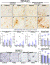
Figure 2.. Striatum - Reactive Astrogliosis and Astrocyte Density.
Representative images from GFAP stained striatum (40x, montage top row) show visible increases in reactive astrogliosis in FXTAS grey (putamen: B,F / caudate: not shown) and white matter (internal capsule: D,H) relative to controls (A,E/C,G). FXTAS subjects showed a significant increase in (I) the proportion of astrocytes with a reactive phenotype of any severity (II-IV) in both striatal grey (putamen and caudate) and white matter (internal capsule), (J) the proportion of severely reactive (IV) astrocytes in striatal white matter, and (K) the severity of GFAP+ fragmentation within striatal white matter (fragment scores represent proportion of field containing dense GFAP+ puncta). (L) Astrocyte density is significantly reduced in FXTAS striatal white matter, but not grey matter, relative to controls. Striatal tissue was also stained with the astrocyte marker ALDH1L1 (M-P), confirming that there is (Q) a significant reduction in astrocyte density in FXTAS cerebellar white matter. Mean ± SE shown, Bonferroni adjusted α=.017. Each datapoint represents a single subject’s score, and all scores represent counts averaged across three images per subject per sub-region). N = 18 (n=9 FXTAS, n=9 control) for caudate and internal capsule. N= 23 for putamen (n=12 FXTAS, n=11 control).
Figure 3.
Figure 3.. Cerebellum - Reactive Astrogliosis and Astrocyte Density.
Representative images from GFAP stained cerebellum (40x, montage top row) shows visible increases in reactive gliosis in FXTAS cerebellar white matter (medullary substance, D,H) relative to controls (C,G), but not in grey matter (granule cell layer: A-B, E-F). FXTAS subjects showed a significant increase in (I) the proportion of astrocytes with a reactive phenotype of any severity (II-IV) in cerebellar white matter but not in grey matter, (J) the proportion of severely reactive (IV) astrocytes in cerebellar white matter only, and (K) the severity of GFAP+ fragmentation within cerebellar white matter only (fragment scores represent proportion of field containing dense GFAP+ puncta). (L) Astrocyte density is significantly reduced in FXTAS cerebellar white matter, but not grey matter, relative to controls. Cerebellar tissue was also stained with the astrocyte marker ALDH1L1 (M-P), confirming that there is (Q) a significant reduction in astrocyte density in FXTAS cerebellar white matter. Mean ± SE shown, Bonferroni adjusted α=.025. All scores represent counts averaged across three images per subject. Each datapoint represents a single subject’s score, and all scores represent counts averaged across three images per subject per sub-region. N = 24 (n=12 FXTAS, n=12 control) for granule cell layer and medullary substance.
Figure 4.
Figure 4.. Perivascular Astrogliosis.
Representative images from GFAP stained cerebellar and striatal white matter. (A) Perivascular astrocyte processes are visible in control cerebellar white matter with (C) a cross-sectional [arrows] and (B) transverse orientation [arrowheads]. Low magnification of FXTAS (D, G) cerebellar and (J) striatal white matter, illustrating (E,F) particularly severe perivascular astrocyte reactivity, as well as reactive perivascular fibers in (H,K) cross sectional and (I,L) transverse orientation.
Figure 5.
Figure 5.. Astrocyte Degeneration in Striatal White Matter.
(A) GFAP stained striatal white matter typically reveals numerous resting astrocytes (B,C) and abundant fibers (D), even in areas of low immunoreactivity (E), as illustrated in control subject. In contrast, FXTAS subjects (F,K) showed a consistent but markedly different appearance with fewer astrocytes, a high proportion of dysmorphic reactive (G,L) and fragmented (H,M) astrocytes, large areas filled with clusters of GFAP+ fragments reminiscent of apoptotic blebbing (I,N). Even in areas with low immunoreactivity, blebbing was typically present, although GFAP+ astrocytes were absent (J,O).
Figure 6.
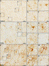
Figure 6.. Astrocyte Degeneration in Cerebellar White Matter.
(A) GFAP stained cerebellar white matter typically reveals numerous resting astrocytes (B,C) and abundant fibers (D), even in areas of low immunoreactivity (E), as illustrated in a control subject. In contrast, FXTAS subjects (F,K) showed a consistent but markedly different appearance with fewer astrocytes, a high proportion of dysmorphic reactive (G,L) and fragmented (H,M) astrocytes, large areas filled with often clustered GFAP+ fragments reminiscent of apoptotic blebbing (I,N). Even in areas with low immunoreactivity, blebbing was typically present, although GFAP+ astrocytes were absent (J,O).
Figure 7.
Figure 7.. Cleaved Caspase 3 positive astrocytes.
Numerous GFAP+ astrocytes in FXTAS (A) striatal and (B) cerebellar white matter were also positive for the apoptosis marker cleaved caspase 3. Representative images from two FXTAS subjects in top rows, control on bottom row. In most astrocytes, cleaved caspase signal was present in both soma and processes. (C) GFAP+ puncta, reminiscent of apoptotic bodies, were widespread in FXTAS white matter and were often positive for cleaved caspase 3. (D) GFAP+ perivascular astrocyte processes were also positive for cleaved caspase 3.

Comment in

References

    1. Hagerman PJ, Greco CM, Hagerman RJ. A cerebellar tremor/ataxia syndrome among fragile X premutation carriers. Cytogenet Genome Res. 2003;100(1–4):206–12. - PubMed
    1. Hagerman RJ, Hagerman P. Fragile X-associated tremor/ataxia syndrome - features, mechanisms and management. Nat Rev Neurol. 2016. Jul;12(7):403–12. - PubMed
    1. Grigsby J, Brega AG, Engle K, et al. Cognitive profile of fragile X premutation carriers with and without fragile X-associated tremor/ataxia syndrome. Neuropsychology. 2008. Jan;22(1):48–60. - PubMed
    1. Hamlin AA, Sukharev D, Campos L, et al. Hypertension in FMR1 premutation males with and without fragile X-associated tremor/ataxia syndrome (FXTAS). Am J Med Genet A. 2012. Jun;158A(6):1304–9. - PMC - PubMed
    1. Hagerman RJ, Coffey SM, Maselli R, et al. Neuropathy as a presenting feature in fragile X-associated tremor/ataxia syndrome. Am J Med Genet A. 2007. Oct 1;143A(19):2256–60. - PubMed

Supplementary concepts